??
| 中文名稱 | 突觸蛋白6抗體 |
| 別????名 | Synaptotagmin 6; Synaptotagmin VI; Synaptotagmin-6; Synaptotagmin6; SynaptotagminVI; Syt 6; Syt VI; Syt6; SYT6_HUMAN; SytVI.?? |
| 研究領域 | 免疫學??神經生物學??信號轉導??轉運蛋白??細胞類型標志物?? |
| 抗體來源 | Rabbit |
| 克隆類型 | Polyclonal |
| 交叉反應 | Human,?Mouse,?Rat,? (predicted: Pig,?Cow,?Horse,?Rabbit,?) |
| 產品應用 | WB=1:500-2000?ELISA=1:500-1000?IHC-P=1:100-500?IHC-F=1:100-500?IF=1:100-500?(石蠟切片需做抗原修復) not yet tested in other applications. optimal dilutions/concentrations should be determined by the end user. |
| 分?子?量 | 57kDa |
| 細胞定位 | 細胞漿?細胞膜? |
| 性????狀 | Liquid |
| 濃????度 | 1mg/ml |
| 免?疫?原 | KLH conjugated synthetic peptide derived from human Synaptotagmin VI (385-430aa):381-510/510? |
| 亞????型 | IgG |
| 純化方法 | affinity purified by Protein A |
| 儲?存?液 | 0.01M TBS(pH7.4) with 1% BSA, 0.03% Proclin300 and 50% Glycerol. |
| 保存條件 | Shipped at 4℃. Store at -20 °C for one year. Avoid repeated freeze/thaw cycles. |
| PubMed | PubMed |
| 產品介紹 | The protein encoded by this gene belongs to the synaptotagmin family. Synaptotagmins share a common domain structure that includes a transmembrane domain and a cytoplasmic region composed of 2 C2 domains, and are involved in calcium-dependent exocytosis of synaptic vesicles. This protein has been shown to be a key component of the secretory machinery involved in acrosomal exocytosis. Alternatively spliced transcript variants have been found for this gene. [provided by RefSeq, Dec 2011] Function: May be involved in Ca(2+)-dependent exocytosis of secretory vesicles through Ca(2+) and phospholipid binding to the C2 domain or may serve as Ca(2+) sensors in the process of vesicular trafficking and exocytosis. May mediate Ca(2+)-regulation of exocytosis in acrosomal reaction in sperm (By similarity). Subunit: Homodimer (isoform 1). Isoform 1 forms heterodimers with SytIII, SytV and SytX. Interacts with STX1A, STX1B and STX2; the interaction is Ca(2+)-dependent. Isoform 2 is not able to form homodimer and heterodimers. Subcellular Location: Cytoplasmic vesicle, secretory vesicle, synaptic vesicle membrane; Single-pass membrane protein. Isoform 1: Membrane; Single-pass membrane protein. Note=Localized predominantly to endoplasmic reticulum (ER) and/or Golgi-like perinuclear compartment. Isoform 2: Cytoplasm, cytosol. Cell membrane; Peripheral membrane protein. Similarity: Belongs to the synaptotagmin family. Contains 2 C2 domains. SWISS: Q5T7P8 Gene ID: 148281 Database links: Entrez Gene: 148281?Human Entrez Gene: 54524?Mouse Entrez Gene: 60565?Rat Omim: 607718?Human SwissProt: Q5T7P8?Human SwissProt: Q9R0N8?Mouse SwissProt: Q62746?Rat Unigene: 370963?Human Unigene: 88818?Mouse Unigene: 48074?Rat Important Note: This product as supplied is intended for research use only, not for use in human, therapeutic or diagnostic applications. ? |
| 產品圖片 | 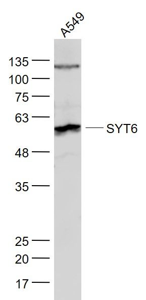 Sample: Sample:A549(Human) Cell Lysate at 30 ug Primary: Anti- SYT6 (bs-4175R) at 1/1000 dilution Secondary: IRDye800CW Goat Anti-Rabbit IgG at 1/20000 dilution Predicted band size: 57 kD Observed band size: 57 kD 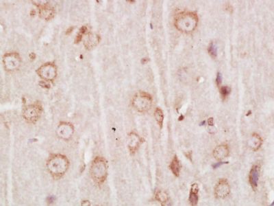 Paraformaldehyde-fixed, paraffin embedded (Rat brain); Antigen retrieval by boiling in sodium citrate buffer (pH6.0) for 15min; Block endogenous peroxidase by 3% hydrogen peroxide for 20 minutes; Blocking buffer (normal goat serum) at 37°C for 30min; Antibody incubation with (SYT6) Polyclonal Antibody, Unconjugated (bs-4175R) at 1:400 overnight at 4°C, followed by a conjugated secondary antibody (sp-0023) for 20 minutes and DAB staining. Paraformaldehyde-fixed, paraffin embedded (Rat brain); Antigen retrieval by boiling in sodium citrate buffer (pH6.0) for 15min; Block endogenous peroxidase by 3% hydrogen peroxide for 20 minutes; Blocking buffer (normal goat serum) at 37°C for 30min; Antibody incubation with (SYT6) Polyclonal Antibody, Unconjugated (bs-4175R) at 1:400 overnight at 4°C, followed by a conjugated secondary antibody (sp-0023) for 20 minutes and DAB staining.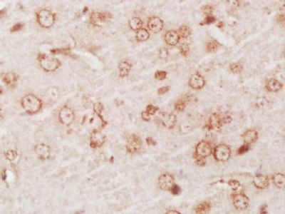 Paraformaldehyde-fixed, paraffin embedded (Mouse brain); Antigen retrieval by boiling in sodium citrate buffer (pH6.0) for 15min; Block endogenous peroxidase by 3% hydrogen peroxide for 20 minutes; Blocking buffer (normal goat serum) at 37°C for 30min; Antibody incubation with (SYT6) Polyclonal Antibody, Unconjugated (bs-4175R) at 1:400 overnight at 4°C, followed by a conjugated secondary antibody (sp-0023) for 20 minutes and DAB staining. Paraformaldehyde-fixed, paraffin embedded (Mouse brain); Antigen retrieval by boiling in sodium citrate buffer (pH6.0) for 15min; Block endogenous peroxidase by 3% hydrogen peroxide for 20 minutes; Blocking buffer (normal goat serum) at 37°C for 30min; Antibody incubation with (SYT6) Polyclonal Antibody, Unconjugated (bs-4175R) at 1:400 overnight at 4°C, followed by a conjugated secondary antibody (sp-0023) for 20 minutes and DAB staining.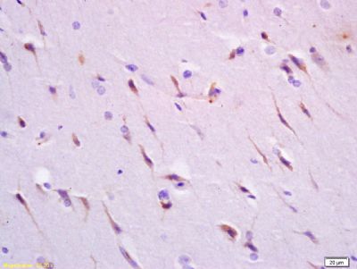 Tissue/cell: human brain tissue; 4% Paraformaldehyde-fixed and paraffin-embedded; Tissue/cell: human brain tissue; 4% Paraformaldehyde-fixed and paraffin-embedded;Antigen retrieval: citrate buffer ( 0.01M, pH 6.0 ), Boiling bathing for 15min; Block endogenous peroxidase by 3% Hydrogen peroxide for 30min; Blocking buffer (normal goat serum,C-0005) at 37℃ for 20 min; Incubation: Anti-SynaptotagminVI Polyclonal Antibody, Unconjugated(bs-4175R) 1:200, overnight at 4°C, followed by conjugation to the secondary antibody(SP-0023) and DAB(C-0010) staining |