??
Entrez Gene: 4133?Human Entrez Gene: 17756?Mouse Entrez Gene: 25595?Rat Omim: 157130?Human SwissProt: P11137?Human SwissProt: P20357?Mouse SwissProt: P15146?Rat Unigene: 368281?Human Unigene: 256966?Mouse Unigene: 10484?Rat
?
中文名稱
磷酸化微管相關(guān)蛋白2抗體
別????名
MAP2(Phospho Ser136); MAP2 (phospho S136); p-MAP2 (phospho S136); MAP2(Phospho S136); MAP2(Phospho-Ser136); p-MAP2(Phospho-Ser136); DKFZp686I2148; Dendrite specific MAP; DKFZp686I2148; MAP 2; MAP-2; MAP2; MAP2_HUMAN; MAP2A; MAP2B; MAP2C; Microtubule associated protein 2; Microtubule-associated protein 2; Mtap 2.??
產(chǎn)品類型
磷酸化抗體?
研究領(lǐng)域
免疫學(xué)??神經(jīng)生物學(xué)??信號轉(zhuǎn)導(dǎo)??干細胞??細胞凋亡??細胞類型標(biāo)志物??細胞骨架??
抗體來源
Rabbit
克隆類型
Polyclonal
交叉反應(yīng)
Human,?Rat,? (predicted: Mouse,?Dog,?Pig,?Cow,?Rabbit,?)
產(chǎn)品應(yīng)用
ELISA=1:500-1000?IHC-P=1:100-500?IHC-F=1:100-500?Flow-Cyt=0.2μg /Test?IF=1:100-500?(石蠟切片需做抗原修復(fù))
not yet tested in other applications.
optimal dilutions/concentrations should be determined by the end user.
分?子?量
70/201kDa
細胞定位
細胞核?細胞漿?
性????狀
Liquid
濃????度
1mg/ml
免?疫?原
KLH conjugated synthesised phosphopeptide derived from human MAP2 around the phosphorylation site of Ser136:PP(p-S)P?
亞????型
IgG
純化方法
affinity purified by Protein A
儲?存?液
0.01M TBS(pH7.4) with 1% BSA, 0.03% Proclin300 and 50% Glycerol.
保存條件
Shipped at 4℃. Store at -20 °C for one year. Avoid repeated freeze/thaw cycles.
PubMed
PubMed
產(chǎn)品介紹
MAP2 is the major microtubule associated protein of brain tissue. There are three forms of MAP2; two are similarily sized with apparent molecular weights of 280 kDa (MAP2a and MAP2b) and the third with a lower molecular weight of 70 kDa (MAP2c). In the newborn rat brain, MAP2b and MAP2c are present, while MAP2a is absent. Between postnatal days 10 and 20, MAP2a appears. At the same time, the level of MAP2c drops by 10-fold. This change happens during the period when dendrite growth is completed and when neurons have reached their mature morphology. MAP2 is degraded by a Cathepsin D-like protease in the brain of aged rats. There is some indication that MAP2 is expressed at higher levels in some types of neurons than in other types. MAP2 is known to promote microtubule assembly and to form side-arms on microtubules. It also interacts with neurofilaments, actin, and other elements of the cytoskeleton.
Function:
The exact function of MAP2 is unknown but MAPs may stabilize the microtubules against depolymerization. They also seem to have a stiffening effect on microtubules.
Subcellular Location:
Cytoplasm, cytoskeleton (Probable).
Post-translational modifications:
Phosphorylated at serine residues in K-X-G-S motifs by MAP/microtubule affinity-regulating kinase (MARK1 or MARK2), causing detachment from microtubules, and their disassembly (By similarity). MAP2A/c is phosphorylated. Isoform MAP2c is phosphorylated by FYN at Tyr-67.
Similarity:
Contains 3 Tau/MAP repeats.
SWISS:
P11137
Gene ID:
4133
Database links:
Important Note:
This product as supplied is intended for research use only, not for use in human, therapeutic or diagnostic applications.
微管相關(guān)蛋白 2(MAP-2)是一種磷蛋白質(zhì),在正常腦組織中,主要存在于神經(jīng)元的胞體、樹突和樹突棘,是腦內(nèi)最豐富的蛋白之一。MAP-2在其調(diào)節(jié)微管的聚合作用和微管的穩(wěn)定性以及對神經(jīng)元軸突和樹突的發(fā)生、延長、穩(wěn)定和突觸可塑性調(diào)節(jié)具有重要作用。
產(chǎn)品圖片
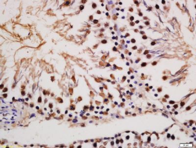 Tissue/cell: rat testis tissue; 4% Paraformaldehyde-fixed and paraffin-embedded;
Antigen retrieval: citrate buffer ( 0.01M, pH 6.0 ), Boiling bathing for 15min; Block endogenous peroxidase by 3% Hydrogen peroxide for 30min; Blocking buffer (normal goat serum,C-0005) at 37℃ for 20 min;
Incubation: Anti-Phospho-MAP2 (Ser136) Polyclonal Antibody, Unconjugated(bs-3259R) 1:200, overnight at 4°C, followed by conjugation to the secondary antibody(SP-0023) and DAB(C-0010) staining
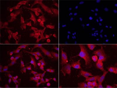 Tissue/cell: human glioma cells, U251;4% Paraformaldehyde-fixed;
Blocking buffer (normal goat serum,C-0005) at 37℃ for 20 min;
Incubation: Anti-Phospho-MAP2(Ser136) Polyclonal Antibody, Unconjugated(bs-3259R) 1:200, overnight at 4°C; The secondary antibody was Goat Anti-Rabbit IgG, Cy3 conjugated (bs-0295G-Cy3)used at 1:200 dilution for 40 minutes at 37°C. DAPI(5ug/ml,blue,C-0033) was used to stain the cell nuclei
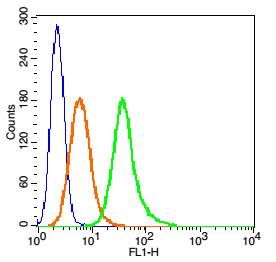 Blank control: RSC96 Cells(blue).
Primary Antibody: Rabbit Anti-hospho-MAP2(Ser136)/FITC Conjugated antibody (bs-3259R-FITC), Dilution: 0.2μg in 100 μL 1X PBS containing 0.5% BSA;
Isotype Control Antibody: Rabbit IgG/FITC(orange) ,used under the same conditions.
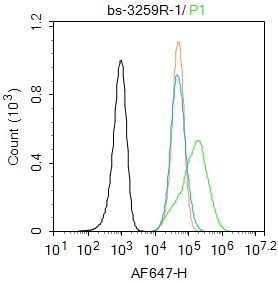 Blank control: SH-SY5Y.
Primary Antibody (green line): Rabbit Anti-Phospho-MAP2 (Ser136) antibody (bs-3259R)
Dilution: 1μg /10^6 cells;
Isotype Control Antibody (orange line): Rabbit IgG .
Secondary Antibody : Goat anti-rabbit IgG-AF647
Dilution: 1μg /test.
Protocol
The cells were fixed with 4% PFA (10min at room temperature)and then permeabilized with 90% ice-cold methanol for 20 min at-20℃.The cells were then incubated in 5%BSA to block non-specific protein-protein interactions for 30 min at room temperature .Cells stained with Primary Antibody for 30 min at room temperature. The secondary antibody used for 40 min at room temperature. Acquisition of 20,000 events was performed.
磷酸化微管相關(guān)蛋白2抗體
作者:上海雅吉生物科技有限公司 2020-10-29T00:00 (訪問量:6267)
上海雅吉生物科技有限公司 商家主頁
地 址: 上海市閔行區(qū)元江路5500號第1幢5658室
聯(lián)系人: 王源
電 話: 15301693058
傳 真: 021-34661275
Email:yajikit@163.com
相關(guān)咨詢
人原代附睪管上皮細胞 (2021-07-08T14:16 瀏覽數(shù):27686)
雅吉生物熱烈慶祝中國建黨100周年 (2021-07-02T09:17 瀏覽數(shù):22537)
15P-1 (小鼠睪丸上皮細胞) (STR鑒定正確) (2021-06-23T09:59 瀏覽數(shù):25343)
客戶細胞培養(yǎng)過程中常見的問題說明 (2021-06-18T09:54 瀏覽數(shù):28622)
免疫熒光鑒定步驟 (2021-06-18T09:54 瀏覽數(shù):29645)
人真皮成纖維細胞的分離培養(yǎng)制備方式 (2021-06-17T09:04 瀏覽數(shù):25835)
原代角質(zhì)形成細胞的分離培養(yǎng)方法 (2021-06-17T09:03 瀏覽數(shù):28750)
鉆石探頭量子顯微鏡可以探索納米微觀世界的奧秘 (2021-06-16T09:24 瀏覽數(shù):27284)
過氧化物酶體活化受體可以加劇腫瘤形成 (2021-06-16T09:23 瀏覽數(shù):25220)
式細胞術(shù)數(shù)據(jù)分析之——圈門技巧 (2021-06-15T10:11 瀏覽數(shù):25511)