?
| 英文名稱 | Lyn |
| 中文名稱 | 膜相關(guān)蛋白酪氨酸激酶Lyn抗體 |
| 別????名 | Hck 2; JTK 8; JTK8; ONCOGENE LYN; Tyrosine protein kinase LYN; V yes 1 Yamaguchi sarcoma viral related oncogene homolog; Yamaguchi sarcoma viral (v yes 1) related oncogene homolog; AA407514; EC 2.7.10.2; FLJ26625; LYN_HUMAN.? |
Entrez Gene: 4067?Human Entrez Gene: 17096?Mouse Entrez Gene: 81515?Rat Omim: 165120?Human SwissProt: P07948?Human SwissProt: P25911?Mouse SwissProt: Q07014?Rat Unigene: 491767?Human Unigene: 317331?Mouse Unigene: 4338?Rat
?
研究領(lǐng)域
免疫學(xué)??細(xì)胞凋亡??激酶和磷酸酶??細(xì)胞表面分子??
抗體來源
Rabbit
克隆類型
Polyclonal
交叉反應(yīng)
Human,?Mouse,?Rat,? (predicted: Dog,?Cow,?Horse,?Rabbit,?Sheep,?)
產(chǎn)品應(yīng)用
WB=1:500-2000?ELISA=1:500-1000?IHC-P=1:100-500?IHC-F=1:100-500?IF=1:100-500?(石蠟切片需做抗原修復(fù))
not yet tested in other applications.
optimal dilutions/concentrations should be determined by the end user.
分?子?量
58kDa
細(xì)胞定位
細(xì)胞核?細(xì)胞漿?細(xì)胞膜?
性????狀
Liquid
濃????度
1mg/ml
免?疫?原
KLH conjugated synthetic peptide derived from human Lyn:201-300/512?
亞????型
IgG
純化方法
affinity purified by Protein A
儲(chǔ)?存?液
0.01M TBS(pH7.4) with 1% BSA, 0.03% Proclin300 and 50% Glycerol.
保存條件
Shipped at 4℃. Store at -20 °C for one year. Avoid repeated freeze/thaw cycles.
PubMed
PubMed
產(chǎn)品介紹
This gene encodes a tyrosine protein kinase, which maybe involved in the regulation of mast cell degranulation, and erythroid differentiation. Alternatively spliced transcript variants encoding different isoforms have been found for this gene. [provided by RefSeq, Jul 2011]
Function:
Non-receptor tyrosine-protein kinase that transmits signals from cell surface receptors and plays an important role in the regulation of innate and adaptive immune responses, hematopoiesis, responses to growth factors and cytokines, integrin signaling, but also responses to DNA damage and genotoxic agents. Functions primarily as negative regulator, but can also function as activator, depending on the context. Required for the initiation of the B-cell response, but also for its down-regulation and termination. Plays an important role in the regulation of B-cell differentiation, proliferation, survival and apoptosis, and is important for immune self-tolerance. Acts downstream of several immune receptors, including the B-cell receptor, CD79A, CD79B, CD5, CD19, CD22, FCER1, FCGR2, FCGR1A, TLR2 and TLR4. Plays a role in the inflammatory response to bacterial lipopolysaccharide. Mediates the responses to cytokines and growth factors in hematopoietic progenitors, platelets, erythrocytes, and in mature myeloid cells, such as dendritic cells, neutrophils and eosinophils. Acts downstream of EPOR, KIT, MPL, the chemokine receptor CXCR4, as well as the receptors for IL3, IL5 and CSF2. Plays an important role in integrin signaling. Regulates cell proliferation, survival, differentiation, migration, adhesion, degranulation, and cytokine release. Down-regulates signaling pathways by phosphorylation of immunoreceptor tyrosine-based inhibitory motifs (ITIM), that then serve as binding sites for phosphatases, such as PTPN6/SHP-1, PTPN11/SHP-2 and INPP5D/SHIP-1, that modulate signaling by dephosphorylation of kinases and their substrates. Phosphorylates LIME1 in response to CD22 activation. Phosphorylates BTK, CBL, CD5, CD19, CD72, CD79A, CD79B, CSF2RB, DOK1, HCLS1, LILRB3/PIR-B, MS4A2/FCER1B, PTK2B/PYK2, SYK and TEC. Promotes phosphorylation of SIRPA, PTPN6/SHP-1, PTPN11/SHP-2 and INPP5D/SHIP-1. Mediates phosphorylation of the BCR-ABL fusion protein. Required for rapid phosphorylation of FER in response to FCER1 activation. Mediates KIT phosphorylation. Acts as an effector of EPOR (erythropoietin receptor) in controlling KIT expression and may play a role in erythroid differentiation during the switch between proliferation and maturation. Depending on the context, activates or inhibits several signaling cascades. Regulates phosphatidylinositol 3-kinase activity and AKT1 activation. Regulates activation of the MAP kinase signaling cascade, including activation of MAP2K1/MEK1, MAPK1/ERK2, MAPK3/ERK1, MAPK8/JNK1 and MAPK9/JNK2. Mediates activation of STAT5A and/or STAT5B. Phosphorylates LPXN on 'Tyr-72'.
Subunit:
Interacts with TEC. Interacts (via SH2 domain) with FLT3 (tyrosine phosphorylated). Interacts with LIME1 and with CD79A upon activation of the B-cell antigen receptor. Interacts with the B-cell receptor complex. Interacts with phosphorylated THEMIS2. Interacts with EPOR. Interacts with MS4A2/FCER1B. Interaction (via the SH2 and SH3 domains) with MUC1 is stimulated by IL7 and the subsequent phosphorylation increases the binding between MUC1 and CTNNB1/beta-catenin. Interacts with Epstein-Barr virus LMP2A. Interacts with Herpes virus saimiri tyrosine kinase interacting protein (Tip). Interacts with ADAM15. Interacts with NDFIP2 and more weakly with NDFIP1. Interacts with FASLG. Interacts with KIT. Interacts with HCLS1. Interacts with FCGR2B. Interacts with FCGR1A; the interaction may be indirect. Interacts with CD19, CD22, CD79A and CD79B. Interacts (via SH3 domain) with PPP1R15A and PDE4A. Interacts with TGFB1I1. Interacts (via SH3 domain) with PIK3R1, the regulatory subunit of phosphatidylinositol 3-kinase; this interaction enhances phosphatidylinositol 3-kinase activity. Interacts with CSF2RB, the common subunit of the IL3, IL5 and CSF2 receptors. Interacts with PAG1; identified in a complex with PAG1 and STAT3. Interacts with ABL1. Interacts with PTPN6/SHP-1. Interacts (via SH3 domain) with SCIMP (via prolin-rich region). Interacts with LPXN (via LD motif 3) and the interaction is induced upon B-cell antigen receptor (BCR) activation.
Subcellular Location:
Cell membrane. Nucleus. Cytoplasm. Cytoplasm, perinuclear region. Golgi apparatus. Note=Accumulates in the nucleus by inhibition of CRM1-mediated nuclear export. Nuclear accumulation is increased by inhibition of its kinase activity. The trafficking from the Golgi apparatus to the plasma membrane occurs in a kinase domain-dependent but kinase activity independent manner and is mediated by exocytic vesicular transport. Detected on plasma membrane lipid rafts.
Tissue Specificity:
Detected in monocytes (at protein level). Detected in placenta, and in fetal brain, lung, liver and kidney. Widely expressed in a variety of organs, tissues, and cell types such as epidermoid, hematopoietic, and neuronal cells. Expressed in primary neuroblastoma tumors.
Post-translational modifications:
Ubiquitinated by CBL, leading to its degradation. Ubiquitination is SH3-dependent.
Autophosphorylated. Phosphorylated on tyrosine residues in response to KIT signaling. Phosphorylation at Tyr-397 is required for optimal activity. Phosphorylation at Tyr-508 inhibits kinase activity. Phosphorylated at Tyr-508 by CSK. Dephosphorylated by PTPRC/CD45. Becomes rapidly phosphorylated upon activation of the B-cell receptor and the immunoglobulin receptor FCGR1A.
DISEASE:
Note=Constitutively phosphorylated and activated in cells from a number of chronic myelogenous leukemia (CML) and acute myeloid leukemia (AML) patients. Mediates phosphorylation of the BCR-ABL fusion protein. Abnormally elevated expression levels or activation of LYN signaling may play a role in survival and proliferation of some types of cancer cells.
Similarity:
Belongs to the protein kinase superfamily. Tyr protein kinase family. SRC subfamily.
Contains 1 protein kinase domain.
Contains 1 SH2 domain.
Contains 1 SH3 domain.
SWISS:
P07948
Gene ID:
4067
Database links:
Important Note:
This product as supplied is intended for research use only, not for use in human, therapeutic or diagnostic applications.
?
產(chǎn)品圖片
 Sample:
SP2/0(Mouse) Cell Lysate at 30 ug
Primary: Anti-Lyn (bs-2906R) at 1/1000 dilution
Secondary: IRDye800CW Goat Anti-Rabbit IgG at 1/20000 dilution
Predicted band size: 58 kD
Observed band size: 58 kD
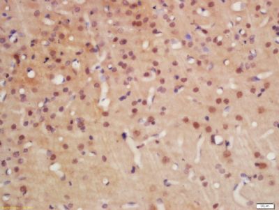 Tissue/cell: rat brain tissue; 4% Paraformaldehyde-fixed and paraffin-embedded;
Antigen retrieval: citrate buffer ( 0.01M, pH 6.0 ), Boiling bathing for 15min; Block endogenous peroxidase by 3% Hydrogen peroxide for 30min; Blocking buffer (normal goat serum,C-0005) at 37℃ for 20 min;
Incubation: Anti-Lyn Polyclonal Antibody, Unconjugated(bs-2906R) 1:200, overnight at 4°C, followed by conjugation to the secondary antibody(SP-0023) and DAB(C-0010) staining
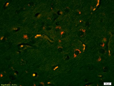 Tissue/cell: human glioma tissue;4% Paraformaldehyde-fixed and paraffin-embedded;
Antigen retrieval: citrate buffer ( 0.01M, pH 6.0 ), Boiling bathing for 15min; Blocking buffer (normal goat serum,C-0005) at 37℃ for 20 min;
Incubation: Anti-Lyn Polyclonal Antibody, PE conjugated (bs-2906R-PE) 1:200, used at 1:200 dilution for 60 minutes at 37°C. Excitation wavelength: 488nm, 555nm; Emission wavelength: 578nm
 Overlay histogram showing Mouse spleen cells stained with bs-2906-PE (red line). The cells were fixed with 1% paraformaldehyde (10 min).The cells were then incubated with the antibody (bs-2906R-PE, 2ug/1x106cells) for 30 min at 22-25°C. Isotype control antibody (black line) was rabbit IgG (2ug/1x106cells) used under the same conditions. Acquisition of events were used for analysis.