??
| 中文名稱 | 造血干細(xì)胞抗原CD133抗體 |
| 別????名 | AC133; Antigen AC133; Hematopoietic stem cell antigen; hProminin; PROM1; Prominin I; Prominin 1; Prominin1; Prominin-1; Prominin like protein 1 precursor; Prominin mouse like 1; prominin1; PROML1; CD133; CORD12; MCDR2; MSTP061; PROML1; RP41; STGD4.?? |
| 研究領(lǐng)域 | 腫瘤??細(xì)胞生物??免疫學(xué)??干細(xì)胞??細(xì)胞類型標(biāo)志物?? |
| 抗體來源 | Rabbit |
| 克隆類型 | Polyclonal |
| 交叉反應(yīng) | Mouse,? (predicted: Human,?Rat,?) |
| 產(chǎn)品應(yīng)用 | WB=1:500-2000?ELISA=1:500-1000?Flow-Cyt=1μg/Test? not yet tested in other applications. optimal dilutions/concentrations should be determined by the end user. |
| 分?子?量 | 95kDa |
| 細(xì)胞定位 | 細(xì)胞膜? |
| 性????狀 | Liquid |
| 濃????度 | 1mg/ml |
| 免?疫?原 | KLH conjugated synthetic peptide derived from human CD133:508-552/865? |
| 亞????型 | IgG |
| 純化方法 | affinity purified by Protein A |
| 儲?存?液 | 0.01M TBS(pH7.4) with 1% BSA, 0.03% Proclin300 and 50% Glycerol. |
| 保存條件 | Shipped at 4℃. Store at -20 °C for one year. Avoid repeated freeze/thaw cycles. |
| PubMed | PubMed |
| 產(chǎn)品介紹 | This gene encodes a pentaspan transmembrane glycoprotein. The protein localizes to membrane protrusions and is often expressed on adult stem cells, where it is thought to function in maintaining stem cell properties by suppressing differentiation. Mutations in this gene have been shown to result in retinitis pigmentosa and Stargardt disease. Expression of this gene is also associated with several types of cancer. This gene is expressed from at least five alternative promoters that are expressed in a tissue-dependent manner. Multiple transcript variants encoding different isoforms have been found for this gene. [provided by RefSeq, Mar 2009] Function: Binds cholesterol in cholesterol-containing plasma membrane microdomains. Proposed to play a role in apical plasma membrane organization of epithelial cells. During early retinal development acts as a key regulator of disk morphogenesis. Involved in regulation of MAPK and Akt signaling pathways. In neuroblastoma cells suppresses cell differentiation such as neurite outgrowth in a RET-dependent manner. Subunit: Interacts with CDHR1 and with actin filaments. Subcellular Location: Cell projection, cilium, photoreceptor outer segment. Isoform 1: Apical cell membrane; Multi-pass membrane protein. Cell projection, microvillus membrane; Multi-pass membrane protein. Note=Found in extracellular membrane particles in various body fluids such as cerebrospinal fluid, saliva, seminal fluid and urine. Tissue Specificity: Isoform 1 is selectively expressed on CD34 hematopoietic stem and progenitor cells in adult and fetal bone marrow, fetal liver, cord blood and adult peripheral blood. Isoform 1 is not detected on other blood cells. Isoform 1 is also expressed in a number of non-lymphoid tissues including retina, pancreas, placenta, kidney, liver, lung, brain and heart. Found in saliva within small membrane particles. Isoform 2 is predominantly expressed in fetal liver, skeletal muscle, kidney, and heart as well as adult pancreas, kidney, liver, lung, and placenta. Isoform 2 is highly expressed in fetal liver, low in bone marrow, and barely detectable in peripheral blood. Isoform 2 is expressed on hematopoietic stem cells and in epidermal basal cells (at protein level). Expressed in adult retina by rod and cone photoreceptor cells (at protein level). Post-translational modifications: Isoform 1 and isoform 2 are glycosylated. DISEASE: Defects in PROM1 are the cause of retinitis pigmentosa type 41 (RP41) [MIM:612095]; also known as retinal degeneration autosomal recessive prominin-related. RP is a retinal dystrophy belonging to the group of pigmentary retinopathies. RP is characterized by retinal pigment deposits visible on fundus examination and primary loss of rod photoreceptor cells followed by secondary loss of cone photoreceptors. Patients typically have night vision blindness and loss of midperipheral visual field. As their condition progresses, they lose their far peripheral visual field and eventually central vision as well. Defects in PROM1 are the cause of cone-rod dystrophy type 12 (CORD12) [MIM:612657]. CORD12 is an inherited retinal dystrophy characterized by retinal pigment deposits visible on fundus examination, predominantly in the macular region, and initial loss of cone photoreceptors followed by rod degeneration. This leads to decreased visual acuity and sensitivity in the central visual field, followed by loss of peripheral vision. Severe loss of vision occurs earlier than in retinitis pigmentosa. Defects in PROM1 are the cause of Stargardt disease type 4 (STGD4) [MIM:603786]. Stargardt disease is the most common hereditary macular degeneration. It is characterized by decreased central vision, atrophy of the macula and underlying retinal pigment epithelium, and frequent presence of prominent flecks in the posterior pole of the retina. Defects in PROM1 are the cause of retinal macular dystrophy type 2 (MCDR2) [MIM:608051]. MCDR2 is a bull's-eye macular dystrophy characterized by bilateral annular atrophy of retinal pigment epithelium at the macula. Similarity: Belongs to the prominin family. SWISS: O43490 Gene ID: 8842 Database links: Entrez Gene: 8842?Human Entrez Gene: 19126?Mouse Entrez Gene: 60357?Rat Omim: 604365?Human SwissProt: O43490?Human SwissProt: O54990?Mouse Unigene: 614734?Human Unigene: 6250?Mouse Unigene: 144589?Rat Important Note: This product as supplied is intended for research use only, not for use in human, therapeutic or diagnostic applications. 一般認(rèn)為,VEGFR2(血管內(nèi)皮生長因子受體2)是HSCs(造血干細(xì)胞)的特異性的表面標(biāo)志。近來經(jīng)研究發(fā)現(xiàn)CD133分子是HSCs(造血干細(xì)胞)特異性標(biāo)志。CD133即AC133,是一個新發(fā)現(xiàn)的HSCs(造血干細(xì)胞)表面標(biāo)志,在HSCs(造血干細(xì)胞)分化成熟過程中,CD133的含量迅速降低。EPCs(血管內(nèi)皮前體細(xì)胞)區(qū)別于成熟內(nèi)皮細(xì)胞的主要標(biāo)志是CD133。 經(jīng)研究發(fā)現(xiàn)內(nèi)皮細(xì)胞不能結(jié)合CD133的抗體。證實分化成熟的內(nèi)皮細(xì)胞不具有CD133。這些說明CD133可以作為EPCs(血管內(nèi)皮前體細(xì)胞)區(qū)別于成熟內(nèi)皮細(xì)胞的一個表面標(biāo)志. |
| 產(chǎn)品圖片 |  Sample: Sample:SP2/0 Cell (Mouse) Lysate at 40 ug Colon carcinoma (Human) Lysate at 40 ug Primary: Anti-CD133 (bs-4770R) at 1/300 dilution Secondary: HRP conjugated Goat-Anti-rabbit IgG (bs-0295G-HRP) at 1/5000 dilution Predicted band size: 95 kD Observed band size: 95/120 kD 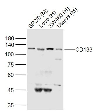 Sample: Sample:Lane 1: SP2/0 (Mouse) Cell Lysate at 30 ug Lane 2: Lovo (Human) Cell Lysate at 30 ug Lane 3: SW480 (Human) Cell Lysate at 30 ug Lane 4: Uterus (Mouse) Lysate at 40 ug Primary: Anti-CD133 (bs-4770R) at 1/300 dilution Secondary: IRDye800CW Goat Anti-Rabbit IgG at 1/20000 dilution Predicted band size: 110 kD Observed band size: 115 kD 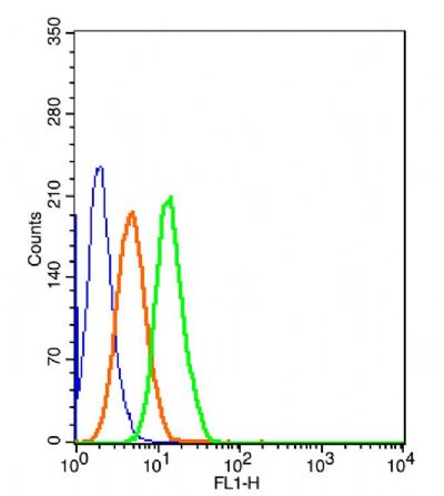 The blue histogram is unstained cells(HepG 2). The blue histogram is unstained cells(HepG 2).The Orange histogram is cells stained with Rabbit IgG/FITC (bs-0295P-FITC) The green histogram is cells stained with Rabbit Anti-CD133/FITC Conjugated antibody (bs-4770R-FITC). Isotype control: Cell lines treated with Rabbit IgG/FITC (bs-0295P-FITC) instead of the primary antibody to confirm that primary antibody binding is 2μg/5μg/1μg in 100μL 1 X PBS containing 0.5% BSA. 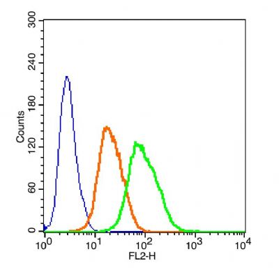 The blue histogram is unstained cells(HepG 2). The blue histogram is unstained cells(HepG 2).The Orange histogram is cells stained with Rabbit IgG/PE (bs-0295P-PE) The green histogram is cells stained with Rabbit Anti-CD133/PE Conjugated antibody (bs-4770R-PE). Isotype control: Cell lines treated with Rabbit IgG/PE (bs-0295P-PE) instead of the primary antibody to confirm that primary antibody binding is specific. 2μg/5μg/10μg in 100μL 1 X PBS containing 0.5% BSA. |
?