血管內皮生長因子C型抗體
產品名稱: 血管內皮生長因子C型抗體
英文名稱: VEGF-C
產品編號: 1586
產品價格: null
產品產地: 上海
品牌商標: 雅吉
更新時間: null
使用范圍: WB ELISA IHC-P IHC-F Flow-Cyt IF
- 聯系人 :
- 地址 : 上海市閔行區元江路5500號第1幢5658室
- 郵編 :
- 所在區域 : 上海
- 電話 : 158****3937 點擊查看
- 傳真 : 點擊查看
- 郵箱 : yajikit@163.com
?
| 中文名稱 | 血管內皮生長因子C型抗體 |
| 別????名 | Vascuoar endothelial growth factor-C; AW228853; Flt4 ligand; Flt4-L; VEGF2; VEGFC; VRP; VEGFC_HUMAN.?? |
?
研究領域細胞生物? 血管內皮細胞??
抗體來源Rabbit
克隆類型Polyclonal
交叉反應Human, Mouse, Rat,?
產品應用WB=1:500-2000 ELISA=1:500-1000 IHC-P=1:100-500 IHC-F=1:100-500 Flow-Cyt=1ug/Test IF=1:100-500 (石蠟切片需做抗原修復)
not yet tested in other applications.
optimal dilutions/concentrations should be determined by the end user.
分 子 量46kDa
細胞定位分泌型蛋白?
性? ? 狀Liquid
濃? ? 度1mg/ml
免 疫 原KLH conjugated synthetic peptide derived from human VEGF-C:321-415/415?
亞? ? 型IgG
純化方法affinity purified by Protein A
儲 存 液0.01M TBS(pH7.4) with 1% BSA, 0.03% Proclin300 and 50% Glycerol.
保存條件Shipped at 4℃. Store at -20 °C for one year. Avoid repeated freeze/thaw cycles.
PubMedPubMed
產品介紹Vascular endothelial growth factors (VEGFs), also known as vasculotropins, are a family of closely related growth factors having a conserved pattern of eight cysteine residues and sharing common VEGF receptors. VEGFs stimulate the proliferation of endothelial cells, induce angiogenesis, promote cell migration, increase vascular permeability, and inhibit apoptosis. The mitogenic activity of VEGFs appears to be mediated by specific VEGF receptors. The target cell specificity of VEGF is restricted to vascular endothelial cells. Vascular Endothelial Growth Factor C (VEGFC) is a member of the VEGF subfamily of PDGF-related growth factors. It is the ligand for Flt4 (VEGFR3) and KDR (VEGFR2). VEGFC binds Flt4 and induces tyrosine autophosphorylation of VEGFR3 and VEGFR2. VEGFC also stimulates the migration of bovine capillary endothelial cells in collagen gel. It is a specific growth factor for the lymphatic vascular system and mediates lymphangiogenesis. VEGFC is abundantly expressed in heart and skeletal muscle. Other tissues such as lung and kidney also express VEGFC.
?
Subunit:
Homodimer; non-covalent and antiparallel.
?
Subcellular Location:
Secreted.
?
Tissue Specificity:
Spleen, lymph node, thymus, appendix, bone marrow, heart, placenta, ovary, skeletal muscle, prostate, testis, colon and small intestine and fetal liver, lung and kidney, but not in peripheral blood lymphocyte.
?
Similarity:
Belongs to the PDGF/VEGF growth factor family.
?
SWISS:
P97953
?
Gene ID:
7424
?
Database links:
Entrez Gene: 7424 Human
?
Entrez Gene: 22341 Mouse
?
Entrez Gene: 114111 Rat
?
Omim: 601528 Human
?
SwissProt: P49767 Human
?
SwissProt: P97953 Mouse
?
SwissProt: O35757 Rat
?
Unigene: 435215 Human
?
Unigene: 1402 Mouse
?
Unigene: 6913 Rat
?
?
?
Important Note:
This product as supplied is intended for research use only, not for use in human, therapeutic or diagnostic applications.
| 產品圖片 |
 Sample:
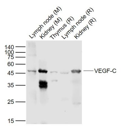
Lane 1: Lymph node (Mouse) Lysate at 40 ug Lane 2: Kidney (Mouse) Lysate at 40 ug Lane 3: Thymus (Rat) Lysate at 40 ug Lane 4: Lymph node (Rat) Lysate at 40 ug Lane 5: Kidney (Rat) Lysate at 40 ug Primary: Anti-VEGF-C (bs-1586R) at 1/1000 dilution Secondary: IRDye800CW Goat Anti-Rabbit IgG at 1/20000 dilution Predicted band size: 46 kD Observed band size: 46 kD 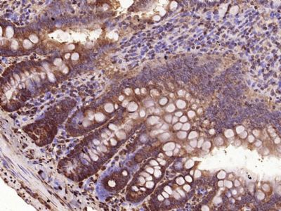 Paraformaldehyde-fixed, paraffin embedded (Rat small intestine); Antigen retrieval by microwave in sodium citrate buffer (pH6.0) ; Block endogenous peroxidase by 3% hydrogen peroxide for 30 minutes; Blocking buffer (3% BSA) at RT for 30min; Antibody incubation with (VEGF-C) Polyclonal Antibody, Unconjugated (bs-1586R) at 1:400 overnight at 4℃, followed by conjugation to the secondary antibody (labeled with HRP)and DAB staining.
 Blank control: HepG2.
Primary Antibody (green line): Rabbit Anti-VEGF-C antibody (bs-1586R) Dilution: 1μg /10^6 cells; Isotype Control Antibody (orange line): Rabbit IgG . Secondary Antibody : Goat anti-rabbit IgG-AF647 Dilution: 1μg /test. Protocol The cells were fixed with 4% PFA (10min at room temperature)and then permeabilized with 0.1% PBST for 20 min at room temperature. The cells were then incubated in 5%BSA to block non-specific protein-protein interactions for 30 min at room temperature .Cells stained with Primary Antibody for 30 min at room temperature. The secondary antibody used for 40 min at room temperature. Acquisition of 20,000 events was performed. 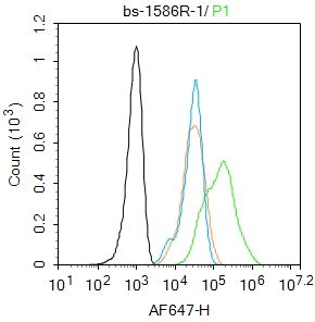 Blank control: HepG2.
Primary Antibody (green line): Rabbit Anti-VEGF-C antibody (bs-1586R) Dilution: 1μg /10^6 cells; Isotype Control Antibody (orange line): Rabbit IgG . Secondary Antibody : Goat anti-rabbit IgG-AF647 Dilution: 1μg /test. Protocol The cells were fixed with 4% PFA (10min at room temperature)and then permeabilized with 0.1% PBST for 20 min at room temperature. The cells were then incubated in 5%BSA to block non-specific protein-protein interactions for 30 min at room temperature .Cells stained with Primary Antibody for 30 min at room temperature. The secondary antibody used for 40 min at room temperature. Acquisition of 20,000 events was performed. |
| ? |