
Cell Counting Kit (CCK-8) CCK-8試劑盒
產品名稱: Cell Counting Kit (CCK-8) CCK-8試劑盒
英文名稱: Cell Counting Kit (CCK-8) CCK-8試劑盒
產品編號: 40203ES
產品價格: 詢價
產品產地: 翌圣生物
品牌商標: Yeasen
更新時間: 2025-11-13T12:42:06
使用范圍: null
- 聯系人 : 李自轉
- 地址 : 上海市浦東新區天雄路166弄一號樓三層南單元
- 郵編 : 200030
- 所在區域 : 上海
- 電話 : 139****5640 點擊查看
- 傳真 : 點擊查看
- 郵箱 : lizizhuan@yeasen.com
- 二維碼 : 點擊查看
Cell Counting Kit-8簡稱CCK-8試劑盒,是一種基于WST-8(化學名:2-(2-甲氧基-4-硝苯基)-3-(4-硝苯基)-5-(2,4-二磺基苯)-2H-四唑單鈉鹽)的廣泛應用于細胞增殖和細胞毒性的快速高靈敏度檢測試劑盒。
WST-8屬于MTT的升級產品,工作原理為:在電子耦合試劑存在的情況下,可以被線粒體內的脫氫酶還原生成高度水溶性的橙黃色的甲臜產物(formazan)。顏色的深淺與細胞的增殖成正比,與細胞毒性成反比。使用酶標儀在450nm波長處測定OD值,間接反映活細胞數量。
CCK-8法應用非常廣泛,如藥物篩選、細胞增殖測定、細胞毒性測定、腫瘤藥敏試驗以及生物因子的活性檢測等。
操作簡單:產品本身是液體,即開即用;甲臜產物是水溶性的,不需要用有機溶劑溶解。
靈敏度高:線性范圍廣,靈敏度高,重復性好。
細胞毒性低:細胞毒性非常低,對后續實驗沒有影響。
穩定性好:培養液中酚紅和血清的存在不會對檢測造成影響。
應用性廣:可用于大規模、高通量樣品檢測。
同類產品性能比較:
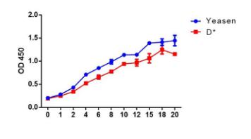
細胞類型:293T人胚腎細胞;培養基:10% FBS 高糖DMEM培養基
孵育時間:3h;細胞數目:1000
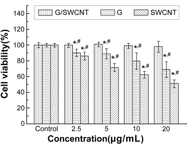
引用已發表文獻數據:
 |
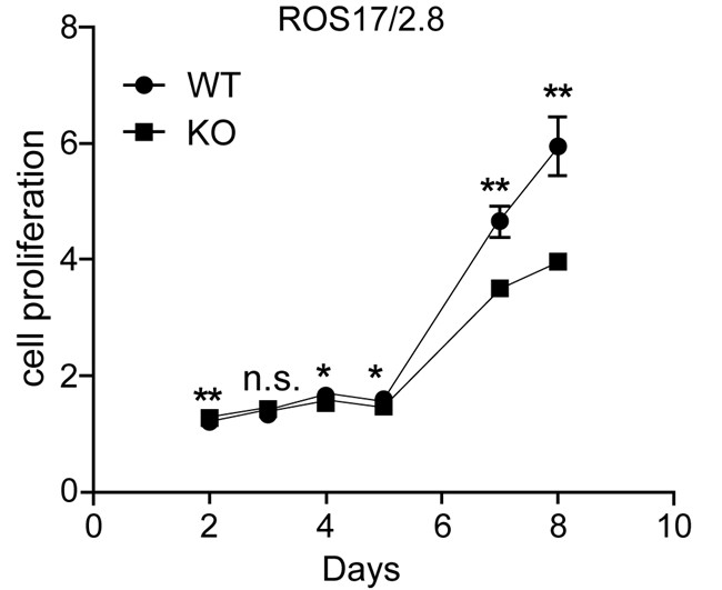 |
| GIST-T1細胞增殖檢測CRTL為空白對照組,Lv-shNC為陰性干擾組,Lv-shOrai1為Orai1敲除組 | ROS17/2.8細胞增殖檢測WT為野生型,KO為BK基因敲除組 |
 |
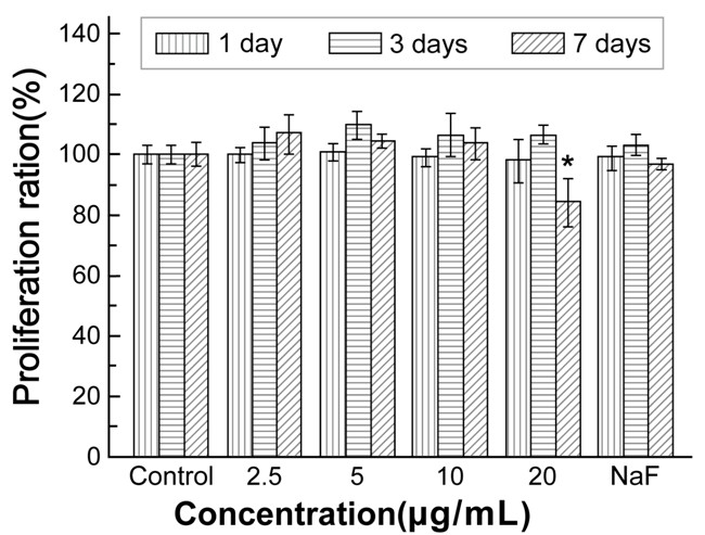 |
| rMSCs細胞活性檢測在2.5,5,10,20ug/mL的濃度 G/SWCNT hybrids, G, and SWCNTs處理下的細胞活性檢測 | rMSCs細胞增殖檢測G/SWCNT hybrids 在2.5,5,10,20ug/mL的濃度下處理1,3和7天細胞活性檢測 |
 |
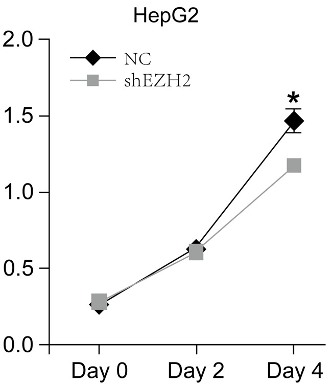 |
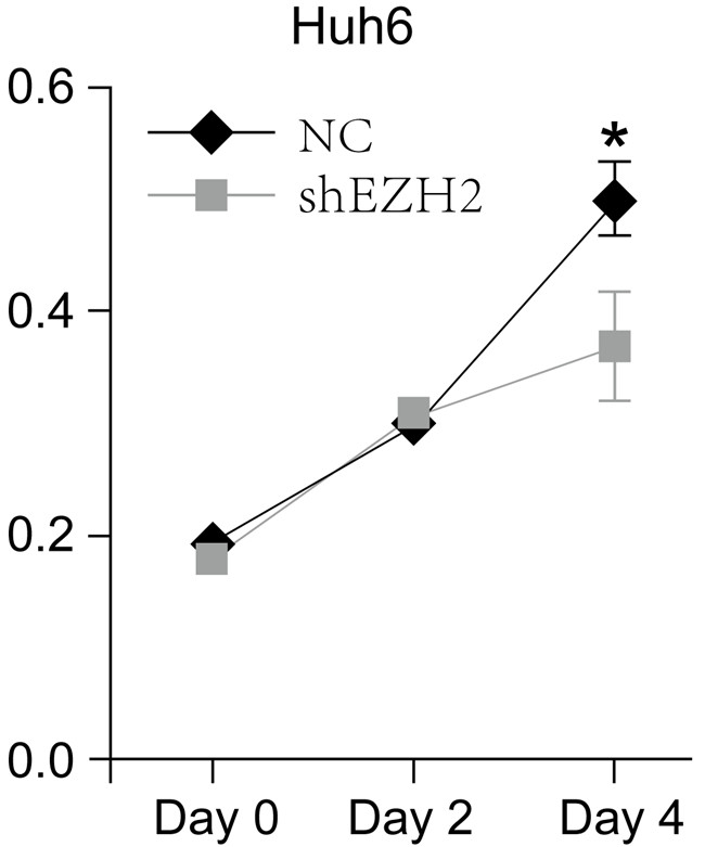
| Huh6細胞干擾EZH2基因和陰性對照組細胞活性檢測 | HepG2細胞干擾EZH2基因和陰性對照組細胞活性檢測 |
 |
 |
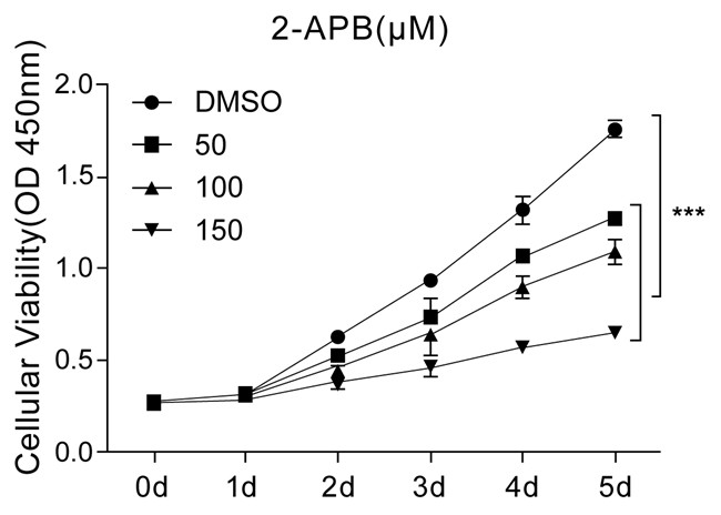
| GIST-T1 細胞活性檢測分別在2-APB 不同濃度0, 50, 100, and 150µM處理下的細胞活力檢測 | GIST-T1 細胞活性檢測分別在SKF-96365不同濃度下0, 0.5, 1.0, and 1.5µM處理下胞活力檢測 |
冰袋運輸,-25~-15“C避光干燥保存,有效期2年。
Q:CCK-8 的保存和穩定性如何?
A:CCK-8 穩定性高,避光條件-20℃有效期 2 年,4℃有效期 1 年。經常使用建議 4℃ 保存,避免反復凍融。CCK 正常顏色為粉紅色,若顏色發生明顯偏差,質量可能有發生變化。
Q:CCK-8 的細胞毒性如何?
A:CCK-8 毒性非常低,因此 CCK-8 檢測完后相同的細胞還能用于其它細胞增殖檢測如結晶紫染色法,中性紅染色法或 DNA 熒光染色法。
Q:CCK-8 會對活細胞進行染色嗎?
A:不會,首先 WST-8 不會進入細胞染色,整個顯色反應都是在培養體系中進行的。另外 WST 和 WST-8 甲臜都屬于高度水溶性組分,反應結束更換新的培養液,即可清除外源組分。因為 CCK-8 是基于 WST-8 能夠被線粒體內的脫氫酶還原生成高度水溶性的橙黃色甲臜產物(Formazan)的原理研制開發的,因而不會對細胞進行染色。
Q:培養基的本底顏色如酚紅會不會影響細胞活性的檢測呢?
A:不會的,培養基中酚紅的吸光度可以在計算時,通過扣除空白孔中本底的吸光度而消去,因此不會對檢測造成影響。
Q:在加入 CCK-8 時可否不更換培養基?
A:一般情況下可以不更換培養基。但是如果培養基里有氧化還原性物質的話,有可能會產生誤差,必須更換其它培養基。
Q:在實驗中OD 值太高或者太低,怎么解決?
A:太高可以縮短加入CCK-8 后的培養時間。例如:可以把加入CCK-8 試劑后的培養時間由 2 小時縮短為 1 小時。此外,可適當減少細胞的數量。太低可以采取 2 個辦法: 1、適當增加細胞數量 2、延長加入CCK-8 試劑后的反應時間。
Q:哪些物質會影響 CCK-8 的實驗測定結果?
A:當培養體系內含有還原性物質存在時會還原 WST-8,從而使 OD 值增加;當有氧化性物質存在時會抑制還原反應進而使 OD 值減小。
Q:做細胞毒性實驗,我的細胞都死了,為什么我加入CCK8試劑后檢測的OD值和對照組OD值差不多?
A:①細胞凋亡或者死亡之后乳酸脫氫酶被釋放到細胞培養液中,可以長期存在,從而導致檢測的OD值偏大。再次說明細胞的活性和OD值并不能完全劃等號。建議客戶在加入CCK8的時候更換新的培養基,確保得到準確的結果。
②加入細胞中的檢測藥物是還原性的,從而導致OD值偏高。建議加入CCK8的時候更換新的培養基。
Q:對照組(培養基+CCK8)的值很高,是為什么呢?
A:可以測一下培養基是否含有氧化還原物質,可能是培養基中的物質引起的。
Q: 細胞不貼壁可以測活性嗎?預培養是必須的嗎?
A: 需要預培養,如果要向保持細胞的最好狀態,建議預培養細胞。如果不做細胞預培養 ,細胞內的脫氫酶可能會不穩定。若不做細胞預培養,需在做標準曲線和檢測時需要統一檢測條件。
Q: CCK-8能否檢測細菌細胞?
A: 可以檢測大腸桿菌,但不能檢測酵母細胞。向100μl E.coli培養液中加入10μl CCK-8溶液,并培養1-4個小時或過夜。
- Qian X, Yi W, Yan W, et al. Cryo-Shocked Tumor-Reprogrammed Sonosensitive Antigen-Presenting Cells Improving Sonoimmunotherapy via T Cells and NK Cells Immunity. Adv Mater. Published online February 16, 2025. doi:10.1002/adma.202413289 (IF:27.4)
- Yao C, Ni Z, Gong C, et al. Rocaglamide enhances NK cell-mediated killing of non-small cell lung cancer cells by inhibiting autophagy[J]. Autophagy, 2018, 14(10): 1831-1844. IF(11.1)
- X Fan, H Cheng, et al. Incorporation of Polycaprolactone to Cyclodextrin-Based Nanocarrier for Potent Gene Delivery. Macromolecular materials and engineering, Volume303, Issue9 ,September 2018.IF(2.777)
- Cheng H, Fan X, et al. Cyclodextrin-Based Star-Like Amphiphilic Cationic Polymer as a Potential Pharmaceutical Carrier in Macrophages. Macromol Rapid Commun. 2018 May 28. IF(4.441)
- Ma Q, Zhang Y, Liang H, et al. EMP3, which is regulated by miR-663a, suppresses gallbladder cancer progression via interference with the MAPK/ERK pathway[J]. Cancer letters, 2018, 430: 97-108.IF(6.491)
- Liu Y, Pan Y F, Xue Y, et al. uPAR promotes tumor-like biologic behaviors of fibroblast-like synoviocytes through PI3K/Akt signaling pathway in patients with rheumatoid arthritis[J]. Cellular & molecular immunology, 2018, 15(2): 171. IF(7.551)
- Shen, C.T., et al., Metformin reduces glycometabolism of papillary thyroid carcinoma in vitro and in vivo. J Mol Endocrinol, 2017. 58(1): p. 15-23.IF(3.297)
- Jiang, J.Z., et al., Metabolic-induced cytotoxicity of diosbulbin B in CYP3A4-expressing cells. Toxicol In Vitro, 2017. 38: p. 59-66.IF(3.105)
- Du Z, et al., MicroRNA31-NDRG3 regulation axes are essential for hepatocellular carcinoma survival and drug resistance. Cancer Biomark, 2017.IF(2.392)
- Li, X., et al., Placental growth factor silencing ameliorates liver fibrosis and angiogenesis and inhibits activation of hepatic stellate cells in a murine model of chronic liver disease. J Cell Mol Med, 2017.(4.302)
- Liu, S., et al., Macrophage infiltration of electrospun polyester fibers. Biomater Sci, 2017.IF(5.831)
- Shi, G., et al., Sox9 facilitates proliferation, differentiation and lipogenesis in primary cultured human sebocytes. J Dermatol Sci, 2017. 85(1): p. 44-50.IF(3.675)
- Liu, Y., et al., uPAR promotes tumor-like biologic behaviors of fibroblast-like synoviocytes through PI3K/Akt signaling pathway in patients with rheumatoid arthritis. Cell Mol Immunol, 2017.IF(7.551)
- Zhang, S., J. Yin and J. Zhong, Chaetocin reactivates the lytic replication of Epstein-Barr virus from latency via reactive oxygen species. Sci China Life Sci, 2017. 60(1): p. 66-71.IF(3.085)
- Yang, Q.L., et al., Sweroside ameliorates alpha-naphthylisothiocyanate-induced cholestatic liver injury in mice by regulating bile acids and suppressing pro-inflammatory responses. Acta Pharmacol Sin, 2016. 37(9): p. 1218-28.IF(3.562)
- Wei, W.J., et al., Propranolol sensitizes thyroid cancer cells to cytotoxic effect of vemurafenib. Oncol Rep, 2016. 36(3): p. 1576-84.IF(2.976)
- Yang, Y., et al., Human CIK Cells Loaded with Au Nanorods as a Theranostic Platform for Targeted Photoacoustic Imaging and Enhanced Immunotherapy and Photothermal Therapy. Nanoscale Res Lett, 2016. 11(1): p. 285.IF(3.125)
- Liu, T., et al., Slowly Delivered Icariin/Allogeneic Bone Marrow-Derived Mesenchymal Stem Cells to Promote the Healing of Calvarial Critical-Size Bone Defects. Stem Cells Int, 2016. 2016: p. 1416047.IF(3.989)
- Zhou, Y., et al., Synergistic anti-liver fibrosis actions of total astragalus saponins and glycyrrhizic acid via TGF-beta1/Smads signaling pathway modulation. J Ethnopharmacol, 2016. 190: p. 83-90.IF(3.115)
- Li, W., et al., MicroRNA-495 regulates starvation-induced autophagy by targeting ATG3. FEBS Lett, 2016. 590(6): p. 726-38.IF(2.999)
- Yang, F., et al., Waltonitone inhibits proliferation of hepatoma cells and tumorigenesis via FXR-miR-22-CCNA2 signaling pathway. Oncotarget, 2016. 7(46): p. 75165-75175.IF(2.656)
- Li, S., et al., DNA Cleavage and Condensation Activities of Mono- and Binuclear Hybrid Complexes and Regulation by Graphene Oxide. Molecules, 2016. 21(7).IF(3.098)
- Li, Z.T., et al., Outer membrane vesicles isolated from two clinical Acinetobacter baumannii strains exhibit different toxicity and proteome characteristics. Microb Pathog, 2015. 81: p. 46-52.IF(2.332)
- Xiao, K., et al., Inactivation of BLU is associated with methylation of Sp1-binding site of BLU promoter in gastric cancer. Int J Oncol, 2015. 47(2): p. 621-31.IF(3.333)
- Sun, Y.P., et al., Synovium fragment-derived cells exhibit characteristics similar to those of dissociated multipotent cells in synovial fluid of the temporomandibular joint. PLoS One, 2014. 9(7): p. e101896.IF(2.766)
- Qiao, N., et al., Haemolytic activity and adjuvant effect of soyasaponins and some of their derivatives on the immune responses to ovalbumin in mice. Int Immunopharmacol, 2014. 18(2): p. 333-9.IF(3.118)