溶酶體相關膜蛋白1抗體
產品名稱: 溶酶體相關膜蛋白1抗體
英文名稱: LAMP1
產品編號: 1970
產品價格: null
產品產地: 上海
品牌商標: 雅吉
更新時間: null
使用范圍: ELISA IHC-P IHC-F Flow-Cyt ICC IF
上海雅吉生物科技有限公司
- 聯系人 :
- 地址 : 上海市閔行區元江路5500號第1幢5658室
- 郵編 :
- 所在區域 : 上海
- 電話 : 158****3937 點擊查看
- 傳真 : 點擊查看
- 郵箱 : yajikit@163.com
?
| 中文名稱 | 溶酶體相關膜蛋白1抗體 |
| 別????名 | CD107 antigen like family member A; CD107 antigen-like family member A; CD107a; CD107a antigen; LAMP 1; LAMP-1; LAMP1_HUMAN; LAMPA; LGP120; lgpA; Lysosomal membrane glycoprotein 120KD; Lysosomal Associated Membrane Protein 1; Lysosome associated membrane glycoprotein 1; Lysosome-associated membrane glycoprotein 1; Lysosome-associated membrane glycoprotein 1; Lysosome-associated membrane protein 1; OTTHUMP00000040663.? |
?
| 研究領域 | 腫瘤??免疫學??神經生物學??信號轉導??干細胞??新陳代謝??細胞膜蛋白?? |
| 抗體來源 | Rabbit |
| 克隆類型 | Polyclonal |
| 交叉反應 | Human,?Mouse,?Rat,? |
| 產品應用 | ELISA=1:500-1000?IHC-P=1:100-500?IHC-F=1:100-500?Flow-Cyt=1μg /test?ICC=1:100-500?IF=1:100-500?(石蠟切片需做抗原修復) not yet tested in other applications. optimal dilutions/concentrations should be determined by the end user. |
| 分?子?量 | 42kDa |
| 細胞定位 | 細胞漿?細胞膜? |
| 性????狀 | Liquid |
| 濃????度 | 1mg/ml |
| 免?疫?原 | KLH conjugated synthetic peptide derived from human LAMP-1:301-417/417? |
| 亞????型 | IgG |
| 純化方法 | affinity purified by Protein A |
| 儲?存?液 | 0.01M TBS(pH7.4) with 1% BSA, 0.03% Proclin300 and 50% Glycerol. |
| 保存條件 | Shipped at 4℃. Store at -20 °C for one year. Avoid repeated freeze/thaw cycles. |
| PubMed | PubMed |
| 產品介紹 | Lysosome associated membrane protein (LAMP1), also known as lgp120 or lgpA, is a type 1 integral membrane protein that is transported from trans Golgi networks to endosomes and then lysosomes. Upon cell activation, LAMP1 transfer to the plasma membrane is dependent on a carboxyl terminal tyrosine based motif (YXXI). Perturbation in the spacing between the tyrosine based motif relative to the membrane abolishes lysosome localization of LAMP1. This mutant protein then cycles between the plasma membrane and the endosome. Cell surface LAMP1 and LAMP2 have been shown to promote adhesion of human peripheral blood mononuclear cells (PBMC) to vascular endothelium, therefore they are possibly involved in the adhesion of PBMCs to the site of inflammation. Function: Presents carbohydrate ligands to selectins. Also mplicated in tumor cell metastasis. Subcellular Location: Cell membrane; Single-pass type I membrane protein. Endosome membrane; Single-pass type I membrane protein. Lysosome membrane; Single-pass type I membrane protein. Note=This protein shuttles between lysosomes, endosomes, and the plasma membrane. Post-translational modifications: O- and N-glycosylated; some of the 18 N-linked glycans are polylactosaminoglycans. Similarity: Belongs to the LAMP family. SWISS: P11279 Gene ID: 3916 Database links: Entrez Gene: 3916?Human Entrez Gene: 16783?Mouse Entrez Gene: 25328?Rat Omim: 153330?Human SwissProt: P11279?Human SwissProt: P11438?Mouse SwissProt: P14562?Rat Unigene: 494419?Human Unigene: 16716?Mouse Unigene: 475822?Mouse Unigene: 40177?Rat Important Note: This product as supplied is intended for research use only, not for use in human, therapeutic or diagnostic applications. CD107a可很好的反映NK細胞活性,其表達水平的上調與NK分泌細胞因子的增多及NK對靶細胞的殺傷作用的增強高度相關,采用流式檢測,CD107a還可鑒定出一群無細胞因子分泌的活性NK細胞。 |
| 產品圖片 | 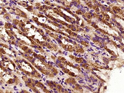 Paraformaldehyde-fixed, paraffin embedded (Rat stomach); Antigen retrieval by boiling in sodium citrate buffer (pH6.0) for 15min; Block endogenous peroxidase by 3% hydrogen peroxide for 20 minutes; Blocking buffer (normal goat serum) at 37°C for 30min; Antibody incubation with (LAMP1) Polyclonal Antibody, Unconjugated (bs-1970R) at 1:400 overnight at 4°C, followed by a conjugated secondary antibody (sp-0023) for 20 minutes and DAB staining. Paraformaldehyde-fixed, paraffin embedded (Rat stomach); Antigen retrieval by boiling in sodium citrate buffer (pH6.0) for 15min; Block endogenous peroxidase by 3% hydrogen peroxide for 20 minutes; Blocking buffer (normal goat serum) at 37°C for 30min; Antibody incubation with (LAMP1) Polyclonal Antibody, Unconjugated (bs-1970R) at 1:400 overnight at 4°C, followed by a conjugated secondary antibody (sp-0023) for 20 minutes and DAB staining.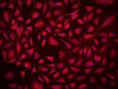 Tissue/cell: A549 cell; 4% Paraformaldehyde-fixed; Triton X-100 at room temperature for 20 min; Blocking buffer (normal goat serum, C-0005) at 37°C for 20 min; Antibody incubation with (LAMP1) Polyclonal Antibody, Unconjugated (bs-1970R) 1:100, 90 minutes at 37°C; followed by a conjugated Goat Anti-Rabbit IgG antibody (bs-0295G-cy3) at 37°C for 90 minutes, DAPI (5ug/ml, blue, C-0033) was used to stain the cell nuclei. Tissue/cell: A549 cell; 4% Paraformaldehyde-fixed; Triton X-100 at room temperature for 20 min; Blocking buffer (normal goat serum, C-0005) at 37°C for 20 min; Antibody incubation with (LAMP1) Polyclonal Antibody, Unconjugated (bs-1970R) 1:100, 90 minutes at 37°C; followed by a conjugated Goat Anti-Rabbit IgG antibody (bs-0295G-cy3) at 37°C for 90 minutes, DAPI (5ug/ml, blue, C-0033) was used to stain the cell nuclei.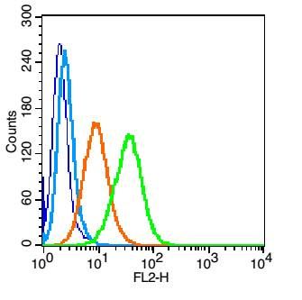 Blank control (blue line): U937 (blue)(fixed with 2% paraformaldehyde for 10 min at room temperature). Blank control (blue line): U937 (blue)(fixed with 2% paraformaldehyde for 10 min at room temperature).Primary Antibody (green line): Rabbit Anti-LAMP1 antibody (bs-1970R); Dilution: 0.2μg /10^6 cells; Isotype Control Antibody (orange line): Rabbit IgG . Secondary Antibody (white blue line): Goat anti-rabbit IgG-PE<; Dilution: 1μg /test. 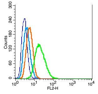 Blank control: RSC96(blue), the cells were fixed with 2% paraformaldehyde (10 min) and then permeabilized with ice-cold 90% methanol for 30 min on ice. Blank control: RSC96(blue), the cells were fixed with 2% paraformaldehyde (10 min) and then permeabilized with ice-cold 90% methanol for 30 min on ice.Isotype Control Antibody: Rabbit IgG(orange) ; Secondary Antibody: Goat anti-rabbit IgG-PE(white blue), Dilution: 1:200 in 1 X PBS containing 0.5% BSA ; Primary Antibody Dilution: 1μg in 100 μL1X PBS containing 0.5% BSA(green). |
?