絲裂原活化蛋白激酶1/ERK 1/2重組兔單克隆抗體
產品名稱: 絲裂原活化蛋白激酶1/ERK 1/2重組兔單克隆抗體
英文名稱: ERK1/2
產品編號: YJ-52924
產品價格: null
產品產地: 上海
品牌商標: 雅吉
更新時間: null
使用范圍: WB, IF, ICC
上海雅吉生物科技有限公司
- 聯系人 :
- 地址 : 上海市閔行區元江路5500號第1幢5658室
- 郵編 :
- 所在區域 : 上海
- 電話 : 158****3937 點擊查看
- 傳真 : 點擊查看
- 郵箱 : yajikit@163.com
?中文名稱 絲裂原活化蛋白激酶1/ERK 1/2重組兔單克隆抗體?別? ? 名 ERK1 + ERK2; ERK 1; ERK 2; ERK-2; ERK1; ERK2; ERT1; ERT2; Extracellular signal regulated kinase 1; Extracellular signal regulated kinase 2; Extracellular signal-regulated kinase 2; HS44KDAP; HUMKER1A; Insulin stimulated MAP2 kinase; MAP kinase 1; MAP kinase 2; MAP kinase isoform p42; MAP kinase isoform p44; MAPK 1; MAPK 2; MAPK 3; MAPK1; MAPK2; MAPK3; MGC20180; Microtubule associated protein 2 kinase; Mitogen activated protein kinase 1; Mitogen activated protein kinase 2; Mitogen activated protein kinase 3; Mitogen-activated protein kinase 1; Mitogen-activated protein kinase 2; MK01_HUMAN; MK03_HUMAN; p38; p40; p41; p41mapk; p42 MAPK; p42-MAPK; p42MAPK; p44 ERK1; p44 MAPK; p44ERK1; p44MAPK; PRKM 1; PRKM 2; PRKM 3; PRKM1; PRKM2; PRKM3; Protein kinase mitogen activated 1; Protein kinase mitogen activated 2; Protein kinase mitogen activated 3; Protein tyrosine kinase ERK 2.? ?研究領域 腫瘤? 細胞生物? 神經生物學? 干細胞? 細胞凋亡? 轉錄調節因子? 激酶和磷酸酶? 細胞骨架? ?抗體來源 Rabbit?克隆類型 Monoclonal?交叉反應 Human, Mouse, Rat,? (predicted: Zebrafish, )?產品應用 WB=1:500-2000 IHC-P=1:50-200 Flow-Cyt=1ug/Test ICC=1:100 (石蠟切片需做抗原修復)not yet tested in other applications.optimal dilutions/concentrations should be determined by the end user.?性? ? 狀 Lyophilized or Liquid?濃? ? 度 1mg/ml?免 疫 原 KLH conjugated synthetic peptide derived from human ERK1/2:??亞? ? 型 IgG?純化方法 affinity purified by Protein A?儲 存 液 0.01M TBS(pH7.4) with 1% BSA, 0.03% Proclin300 and 50% Glycerol.?保存條件 Store at -20 °C for one year. Avoid repeated freeze/thaw cycles.?PubMed PubMed?產品介紹 The protein encoded by this gene is a member of the MAPkinase family. MAP kinases, also known as extracellularsignal-regulated kinases (ERKs), act in a signaling cascade thatregulates various cellular processes such as proliferation,differentiation, and cell cycle progression in response to avariety of extracellular signals. This kinase is activated byupstream kinases, resulting in its translocation to the nucleuswhere it phosphorylates nuclear targets. Alternatively s
產品圖片
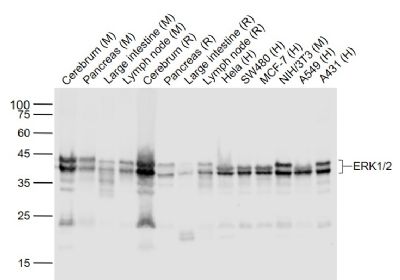
Lane 1: Cerebrum (Mouse) Lysate at 40 ug
Lane 2: Pancreas (Mouse) Lysate at 40 ug
Lane 3: Large intestine (Mouse) Lysate at 40 ug
Lane 4: Lymph node (Mouse) Lysate at 40 ug
Lane 5: Cerebrum (Rat) Lysate at 40 ug
Lane 6: Pancreas (Rat) Lysate at 40 ug
Lane 7: Large intestine (Rat) Lysate at 40 ug
Lane 8: Lymph node (Rat) Lysate at 40 ug
Lane 9: Hela (Human) Cell Lysate at 30 ug
Lane 10: SW480 (Human) Cell Lysate at 30 ug
Lane 11: MCF-7 (Human) Cell Lysate at 30 ug
Lane 12: NIH/3T3 (Mouse) Cell Lysate at 30 ug
Lane 13: A549 (Human) Cell Lysate at 30 ug
Lane 14: A431 (Human) Cell Lysate at 30 ug
Primary: Anti- ERK1/2 (bsm-52259R) at 1/1000 dilution
Secondary: IRDye800CW Goat Anti-Rabbit IgG at 1/20000 dilution
Predicted band size: 44/42 kD
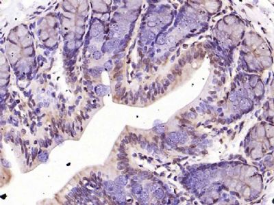
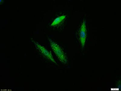
Observed band size: 44/42 kDParaformaldehyde-fixed, paraffin embedded (mouse brain); Antigen retrieval by boiling in sodium citrate buffer (pH6.0) for 15min; Block endogenous peroxidase by 3% hydrogen peroxide for 20 minutes; Blocking buffer (normal goat serum) at 37°C for 30min; Antibody incubation with (ERK1 2) Monoclonal Antibody, Unconjugated (bsm-52259R) at 1:200 overnight at 4°C, followed by operating according to SP Kit(Rabbit) (sp-0023) instructionsand DAB staining.Paraformaldehyde-fixed, paraffin embedded (rat brain); Antigen retrieval by boiling in sodium citrate buffer (pH6.0) for 15min; Block endogenous peroxidase by 3% hydrogen peroxide for 20 minutes; Blocking buffer (normal goat serum) at 37°C for 30min; Antibody incubation with (ERK1 2) Monoclonal Antibody, Unconjugated (bsm-52259R) at 1:200 overnight at 4°C, followed by operating according to SP Kit(Rabbit) (sp-0023) instructionsand DAB staining.Paraformaldehyde-fixed, paraffin embedded (rat colon); Antigen retrieval by boiling in sodium citrate buffer (pH6.0) for 15min; Block endogenous peroxidase by 3% hydrogen peroxide for 20 minutes; Blocking buffer (normal goat serum) at 37°C for 30min; Antibody incubation with (ERK1 2) Monoclonal Antibody, Unconjugated (bsm-52259R) at 1:200 overnight at 4°C, followed by operating according to SP Kit(Rabbit) (sp-0023) instructionsand DAB staining.Tissue/cell:A549 cell;4% Paraformaldehyde-fixed;Triton X-100 at room temperature for 20 min; Blocking buffer (normal goat serum,C-0005) at 37°C for 20 min; Antibody incubation with (ERK1/2) monoclonal Antibody, Unconjugated (bsm-52259R) 1:100, 90 minutes at 37°C; followed by a FITC conjugated Goat Anti-Rabbit IgG antibody at 37°C for 90 minutes, DAPI (blue, C02-04002) was used to stain the cell nuclei.Blank control: Hela.
Primary Antibody (green line): Rabbit Anti-ERK1/2 antibody (bsm-52259R)
Dilution: 1μg /10^6 cells;
Isotype Control Antibody (orange line): Rabbit IgG .
Secondary Antibody : Goat anti-rabbit IgG-AF647
Dilution: 1μg /test.
Protocol
The cells were fixed with 4% PFA (10min at room temperature)and then permeabilized with 90% ice-cold methanol for 20 min at -20℃. The cells were then incubated in 5%BSA to block non-specific protein-protein interactions for 30 min at room temperature .Cells stained with Primary Antibody for 30 min at room temperature. The secondary antibody used for 40 min at room temperature. Acquisition of 20,000 events was performed.
? ?