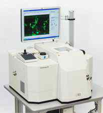
TAXIScan-FL熒光細(xì)胞動(dòng)態(tài)分析系統(tǒng)
產(chǎn)品名稱: TAXIScan-FL熒光細(xì)胞動(dòng)態(tài)分析系統(tǒng)
英文名稱: Fluorescent cell mobility analysis device TAXIScan-FL
產(chǎn)品編號(hào): TAXIScan-FL
產(chǎn)品價(jià)格: 0
產(chǎn)品產(chǎn)地: 日本
品牌商標(biāo): effectorcell
更新時(shí)間: null
使用范圍: null
- 聯(lián)系人 :
- 地址 : 北京市海淀區(qū)西三旗上奧世紀(jì)中心A座9層906
- 郵編 :
- 所在區(qū)域 : 北京
- 電話 : 186****1725 點(diǎn)擊查看
- 傳真 : 點(diǎn)擊查看
- 郵箱 : 787852745@qq.com
制造商(Manufacturer):日本ECI株式會(huì)社(ECI Inc., Japan )
主要技術(shù)指標(biāo)(Main technical indicators):
物鏡:10×20×40×100×(Objective lens: 10×20×40×100×)
熒光濾塊:B/G/R (Fluorescent filter block: B/G/R)
樣品量:≤100個(gè)細(xì)胞(Sample amount: 100 or less cells)
溫度控制:室溫+ 3℃~40℃(Holder temperature control: room temperature+ 3℃~40℃)
硅基底芯片:通道深度4μm,5μm ,6μm,8μm(Chip terrace depth:4, 5, 6, or 8μm)
12個(gè)獨(dú)立通道,可同時(shí)進(jìn)行12例試驗(yàn)(12 channels, up to 12 concurrent assays)
自動(dòng)聚焦系統(tǒng)(Autofocus system)
?
制造商(Manufacturer):日本ECI株式會(huì)社(ECI Inc., Japan )
主要技術(shù)指標(biāo)(Main technical indicators):
物鏡:10×20×40×100×(Objective lens: 10×20×40×100×)
熒光濾塊:B/G/R (Fluorescent filter block: B/G/R)
樣品量:≤100個(gè)細(xì)胞(Sample amount: 100 or less cells)
溫度控制:室溫+ 3℃~40℃(Holder temperature control: room temperature+ 3℃~40℃)
硅基底芯片:通道深度4μm,5μm ,6μm,8μm(Chip terrace depth:4, 5, 6, or 8μm)
12個(gè)獨(dú)立通道,可同時(shí)進(jìn)行12例試驗(yàn)(12 channels, up to 12 concurrent assays)
自動(dòng)聚焦系統(tǒng)(Autofocus system)
動(dòng)態(tài)影像實(shí)時(shí)記錄 (Data store as movie image file)
計(jì)算機(jī)分析系統(tǒng),包含濃度梯度的精確測(cè)量,自動(dòng)統(tǒng)計(jì)細(xì)胞數(shù)量,細(xì)胞形態(tài)變化、遷移速度、遷移方向等統(tǒng)計(jì)學(xué)分析。
主要功能(Main function):硅基底芯片,其上嵌刻的水平通道可形成化學(xué)趨化因子濃度梯度,用于測(cè)定濃度梯度依賴細(xì)胞的功能,如趨化,脫顆粒。水平通道的深度小于懸浮細(xì)胞的直徑,其內(nèi)可觀測(cè)細(xì)胞形態(tài)學(xué)變化和增殖遷移過(guò)程。細(xì)胞趨化分析不僅包括中性粒細(xì)胞、嗜酸性粒細(xì)胞、單核細(xì)胞、淋巴細(xì)胞等外周血白細(xì)胞,也包括各種癌細(xì)胞和培養(yǎng)細(xì)胞,如平滑肌細(xì)胞、內(nèi)皮細(xì)胞、神經(jīng)細(xì)胞、干細(xì)胞等。還可分析蛋白質(zhì)及細(xì)胞相互作用、細(xì)胞信號(hào)轉(zhuǎn)導(dǎo)、細(xì)胞骨架、鈣流入、活性氧代謝等。可應(yīng)用于趨化因子及藥物篩選、炎癥、過(guò)敏反應(yīng)、腫瘤、神經(jīng)、免疫、心血管、干細(xì)胞等方面的研究。
?
日本ECI株式會(huì)社細(xì)胞動(dòng)態(tài)可視化系統(tǒng)設(shè)備TAXIScan-FL,是全新光學(xué)動(dòng)態(tài)成像與活體細(xì)胞處理技術(shù)的完美結(jié)合,本設(shè)備采用專利TAXIScan技術(shù),具有獨(dú)立知識(shí)產(chǎn)權(quán),其核心部件為硅基底芯片,其上嵌刻的水平通道可形成化學(xué)趨化因子濃度梯度;水平通道的深度精度小于懸浮細(xì)胞的直徑,可精確到微米級(jí)別,其內(nèi)可觀測(cè)細(xì)胞形態(tài)學(xué)變化和增值遷移過(guò)程;成像部件冷光CCD相機(jī)定位于觀測(cè)平面以下,配有高性能透鏡和同軸反照明裝置;基于以上的革命性技術(shù)使實(shí)驗(yàn)只需100個(gè)甚至更少的細(xì)胞樣本;根據(jù)實(shí)驗(yàn)具體要求自定義設(shè)置實(shí)驗(yàn)條件參數(shù)。
|
Fluorescent filter block capacity |
Up to 5 blocks |
|
Applicable fluorescent molecules |
Most commercial fluorphores |
|
Bright field optical system |
Reflection plane image system |
|
Objective lens |
10X 20X 40X and 100X |
|
Focus system |
Autofocus compatible |
|
Number of channels |
12 |
|
Holder temperature control |
Room temperature + 3℃~40℃ |
|
Light source |
Mercury lamp |
|
Camera |
Monochrome cooled CCD camera |
|
Equipment control and data analyzing software |
NIS-elements (manufactured by Nikon ) |
|
Main body dimension |
Width 900mm X depth 750mm X height(excluding monitor)1100mm |
|
Rated voltage |
AC 100V 50/60Hz |
|
Wattage |
600VA |

TAXIScan is ECI's unique technology to form chemoattractant concentration gradient in a small space so as to observe cellular chemotaxis movement with good repeatability. Existing model of EZ-TAXIScan with a mere 10X objective lens, limited conventional bright-field observation and up to 6 sample simultaneous assays is appreciated well for its easy use of TAXIScan functions.
Newly introduced TAXIScan-FL makes fluorescent observation possible. The equipment is mounted with up to 40X objective lens and bright field observation system with the use of differential interferometry. The number of simultaneous assay samples is increased up to 12 so that higher analysis with enhanced throughput can be anticipated.

![]()
|
[NEW]Chemotactic Cell Movies are now available |
|
 |
Click the picture |
- The equipment enables fluorescent observation for various types of cell performing chemotaxis movement. Mounted with high-power objective lens (up to 40X)and differential interferometry optical system enable clear image acquisition at bright field observation. The employment of autofocus mechanism also enables stable focus for extended period to time.

- Simultaneous observation of transient increase (in fluorescent strength) of intracellular calcium concentration and chemotaxis movement were performed using cells stained with fluorescent calcium indicator (Fluo-3) (cell: human monocyte, chemoattractant: hMIP-1a)

- EZ-TAXIScan made observation of degranulation phenomena in mast cell possible. In contrast, TAXIScan-FL enables bright field observation in much higher magnification and differencial interferometry. By doing fluorescent straining for mast cells (blue : nucleus, green : calcium concentration, orange : degranulation), it becomes possible to observe transient calcium concentration increase in each cell before degranulation, etc. more in detail.

- Measurement record

- Main specification
