CST PathScan®抗體芯片試劑盒
產品名稱: CST PathScan®抗體芯片試劑盒
英文名稱: PathScan® EGFR Signaling Antibody Array Kit (Chemiluminescent Readout)
產品編號: 12622S
產品價格: null
產品產地: 美國
品牌商標: CST
更新時間: null
使用范圍:
- 聯系人 :
- 地址 : 深圳市寶安區西鄉寶民二路賢基大廈4E
- 郵編 :
- 所在區域 : 廣東
- 電話 : 133****4454 點擊查看
- 傳真 : 點擊查看
- 郵箱 : 1484332550@qq.com
?PathScan?抗體芯片試劑盒是一種基于玻片的ELISA檢測方法,為在多種規格中研究關鍵信號通路節點的信號轉導而設計。
每個試劑盒提供了兩張玻片;每張玻片包含16個分析,每個分析中點了許多抗體。接下來使用檢測抗體混合物來檢測目標蛋白。每個分析中還包含了陽性和陰性對照。
試劑盒分為化學發光檢測和熒光檢測兩種。化學發光檢測適用于膠片,不需要使用其他儀器,而熒光值讀數可以在熒光成像儀中獲取。
試劑盒使用最高質量的抗體進行生產和優化,為您提供了最好的特異性和靈敏性。
試劑盒可以分析小體積裂解物中的多種蛋白,節省寶貴的時間和試劑。
化學發光檢測是一種方便簡單的檢測方法,不需要專門的儀器。
技術支持由CST公司開發和生產這個產品并最了解它們的分子檢測組提供。
12622???? PathScan? EGFR Signaling Antibody Array Kit (Chemiluminescent Readout)
12785???? PathScan? EGFR Signaling Antibody Array Kit (Fluorescent Readout)
12856???? PathScan? Stress and Apoptosis Signaling Antibody Array Kit (Chemiluminescent Readout)
12923???? PathScan? Stress and Apoptosis Signaling Antibody Array Kit (Fluorescent Readout)
13047???? PathScan? Th1/Th2/Th17 Cytokine Antibody Array Kit (Chemiluminescent Readout)
13124???? PathScan? Th1/Th2/Th17 Cytokine Antibody Array Kit (Fluorescent Readout)
13788???? PathScan? Immune Cell Signaling Antibody Array Kit (Fluorescent Readout)
13792???? PathScan? Immune Cell Signaling Antibody Array Kit (Chemiluminescent Readout)
14471???? PathScan? Intracellular Signaling Membrane Array Kit (Chemiluminescent Readout)
14821???? PathScan? Cancer Phenotype Antibody Array Kit (Chemiluminescent Readout)
14822???? PathScan? Cancer Phenotype Antibody Array Kit (Fluorescent Readout)
7949?????? PathScan? RTK Signaling Antibody Array Kit (Fluorescent Readout)
7982?????? PathScan? RTK Signaling Antibody Array Kit (Chemiluminescent Readout)
9474?????? PathScan? Akt Signaling Antibody Array Kit (Chemiluminescent Readout)
?
9700?????? PathScan? Akt Signaling Antibody Array Kit (Fluorescent Readout)
| Array Slides - EGFR Signaling Array Kit? | 2 ea | ? | ? | ? | ? | ? |
| Detection Ab Cocktail A(10X) - EGFR Signaling Array Kit | 150 μl | ? | ? | ? | ? | ? |
| Detection Ab Cocktail B(10X) - EGFR Signaling Array Kit | 150 μl | ? | ? | ? | ? | ? |
| 16-Well Gasket | 2 ea | ? | ? | ? | ? | ? |
| 20X LumiGLO?Reagent and 20X Peroxide #7003 | 5 ml each | ? | ? | ? | ? | ? |
| HRP-Linked Streptavidin (10X) | 300 μl | ? | ? | ? | ? | ? |
| Cell Lysis Buffer (10X) #9803 | 15 ml | ? | ? | ? | ? | ? |
| 20X Array Wash Buffer | 15 ml | ? | ? | ? | ? | ? |
| Sealing Tape | 2 sheets | ? | ? | ? | ? | ? |
| Array Blocking Buffer | 5 ml | ? | ? | ? | ? | ? |
| Chemiluminescent Development Folder | 2 | ? | ? | ? | ? | ? |
| Array Diluent Buffer | 15 ml | ? | ? | ? | ? | ? |
Specificity / Sensitivity
?
PathScan??EGFR Signaling Antibody Array Kit (Chemiluminescent Readout) detects the target proteins as specified on the Array Target Map. No substantial cross-reactivity has been observed between targets. This kit is optimized for cell lysates diluted to a total protein concentration between 0.2 and 1 mg/ml (see kit protocol). All capture antibodies have been validated for human and mouse-derived samples.
?
Description
?
The PathScan??EGFR Signaling Antibody Array Kit (Chemiluminescent Readout) uses glass slides as the planar surface and is based upon the sandwich immunoassay principle. The array kit allows for the simultaneous detection of phosphorylated EGFR, HER2, c-Met on distinct sites as well as a number of key signaling nodes found downstream of these receptor tyrosine kinases (RTKs). Target-specific capture antibodies have been spotted in duplicate onto nitrocellulose-coated glass slides. Each kit contains two slides allowing for the interrogation of 16 different samples. To improve assay performance the content of this array is split between two sub-arrays. The pads on left-hand side of each slide belong to sub-array A while the pads on the right-hand side of each slide belong to sub-array B. Cell lysates are incubated on the slide followed by a biotinylated detection antibody cocktail A or cocktail B (each cocktail for the corresponding sub-array). Streptavidin-conjugated HRP and LumiGLO??Reagent are then used to visualize the bound detection antibody by chemiluminescence. An image of the slide can be captured with either a digital imaging system or standard chemiluminescent film. The image can be analyzed visually or the spot intensities quantified using array analysis software.
?
?
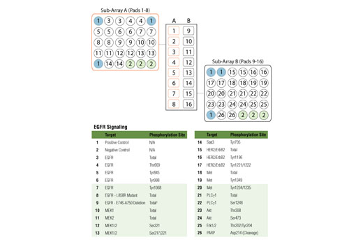
Figure 1. Target map of the PathScan??EGFR Signaling Antibody Array Kit (Chemiluminescent Readout) #12622. ? A reduction in a signal associated with E746-A750 deletion mutant was observed after treatment of cells with the small molecule inhibitors Gefitinib #4765 and Erlotinib #5083.
?
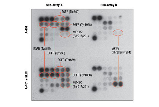
Figure 2. A-431 cells were grown to 90% confluency and then serum-starved overnight. Cells were stimulated with Human Epidermal Growth Factor (hEGF) #8916 (100 ng/ml, 5 min). Cell extracts were prepared and analyzed using the PathScan??EGFR Signaling Antibody Array Kit (Chemiluminescent Readout) #12622. Images were acquired by briefly exposing the slide to standard chemiluminescent film.
?
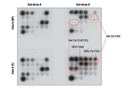
Figure 3. Calu-3 cells were grown to 90% confluency and then lyzed using a buffer containing (P) or devoid of (NP) phosphatase inhibitors. Cell extracts were prepared and analyzed using the PathScan??EGFR Signaling Antibody Array Kit (Chemiluminescent Readout) #12622. Images were acquired by briefly exposing the slide to standard chemiluminescent film.
?
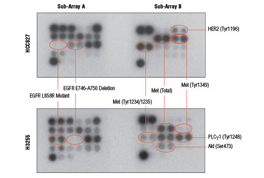
Figure 4. HCC827 and H3255 are two non-small cell lung cancer (NSCLC) cell lines carrying two different gefitinib-sensitive mutants of EGFR: E746-A750 deletion in exon 19 and L858R point mutation, respectively. Cell extracts were prepared and analyzed using the PathScan??EGFR Signaling Antibody Array Kit (Chemiluminescent Readout) #12622. Images were acquired by briefly exposing the slide to standard chemiluminescent film.
Background
?
?
The Epidermal Growth Factor Receptor (EGFR) is a receptor tyrosine kinase (RTK) that constitutes an important disease driver, as well as a validated drug target. The potency of EGFR in driving tumorigenesis can be attributed to its pleiotropic intracellular signaling. Activated EGFR initiates a wide range of signaling modules and switches such as the Ras-Erk/MAP kinase, Akt, Src, Stat, and PKC. Two of the most common EGFR mutations occurring in lung cancer are the E746-A750 deletion and L858R point mutation. This array utilizes unique antibodies made by Cell Signaling Technology that are sensitive to each of these EGFR mutants, allowing specific target detection in cell extracts.
EGFR can interact and heterodimerize with other RTKs. HER2 (also known as ErbB2) is an oncogenic RTK belonging to the EGFR/HER family of RTKs and is an important heterodimerization partner of all HER family members. Another prominent heterodimerization partner of EGFR is c-Met. c-Met is an RTK serving as a receptor for the hepatocyte growth factor (HGF). c-Met can induce cell scattering, migration, and invasion. It has been shown that c-Met is responsible for some cases of tumor resistance to EGFR-targeted therapies and is a contributing factor to tumor metastasis.
PLCγ is a phosphoinositide-specific phospholipase. EGFR can activate PLCγ that, in turn, hydrolyzes phosphoinositide phospholipids residing within the inner leaflet of the plasma membrane. This hydrolysis generates two important second messengers: inositol 1,4,5-triphosphate (IP3) and diacylglycerol (DAG). IP3 causes calcium mobilization from intracellular storage pools, while DAG (together with calcium) activates PKC. MEK1 is a dual-specificity protein kinase and serves as the MAP kinase kinase for Erk1 and Erk2. Upon EGFR activation, MEK1 is phosphorylated by Raf and, in turn, phosphorylates the Erk kinases at Thr202 and Tyr204, leading to their activation. Activated Erk MAP kinase is a major signaling node with a multitude of substrates and primarily transmits growth and proliferation signals. Akt is another important protein kinase downstream of EGFR. Akt is activated by many RTKs and has a large number of intracellular substrates. Akt generates anabolic growth and survival signals. Stat3 is activated in response to EGFR stimulation, as well as in response to activation of a variety of cytokine receptors. Stat3 is a well-established oncogene that is also a transcription factor.
?
The oncogenic signals generated by activated EGFR are a focus of intense drug discovery efforts. It has become clear that in many cases a single agent inhibiting only one target is unable to cause tumor cell death?in vivo. To monitor the blockade of EGFR signals alongside markers of cell death, cleaved PARP is included in this array. PARP is an enzyme involved in DNA repair. As a part of the apoptotic process, PARP is irreversibly inactivated by endoproteolytic cleavage executed by activated cell death proteases, such as caspase-3 and caspasae-7.
12785 PathScan? EGFR Signaling Antibody Array Kit (Fluorescent Readout)
12856 PathScan? Stress and Apoptosis Signaling Antibody Array Kit (Chemiluminescent Readout)
12923 PathScan? Stress and Apoptosis Signaling Antibody Array Kit (Fluorescent Readout)
13047 PathScan? Th1/Th2/Th17 Cytokine Antibody Array Kit (Chemiluminescent Readout)
13124 PathScan? Th1/Th2/Th17 Cytokine Antibody Array Kit (Fluorescent Readout)
7323 PathScan? Intracellular Signaling Array Kit (Chemiluminescent Readout)
7744 PathScan? Intracellular Signaling Array Kit (Fluorescent Readout)
7949 PathScan? RTK Signaling Antibody Array Kit (Fluorescent Readout)
7982 PathScan? RTK Signaling Antibody Array Kit (Chemiluminescent Readout)
9474 PathScan? Akt Signaling Antibody Array Kit (Chemiluminescent Readout)
?
9700 PathScan? Akt Signaling Antibody Array Kit (Fluorescent Readout)