美國Optofluidic激光拉曼鑷子
產(chǎn)品名稱: 美國Optofluidic激光拉曼鑷子
英文名稱: NanoTweezer Raman system
產(chǎn)品編號: NanoTweezer Raman
產(chǎn)品價格: 0
產(chǎn)品產(chǎn)地: 美國
品牌商標: NanoTweezer
更新時間: null
使用范圍: null
- 聯(lián)系人 :
- 地址 : 北京市海淀區(qū)西三旗上奧世紀中心A座9層906
- 郵編 :
- 所在區(qū)域 : 北京
- 電話 : 186****1725 點擊查看
- 傳真 : 點擊查看
- 郵箱 : 787852745@qq.com
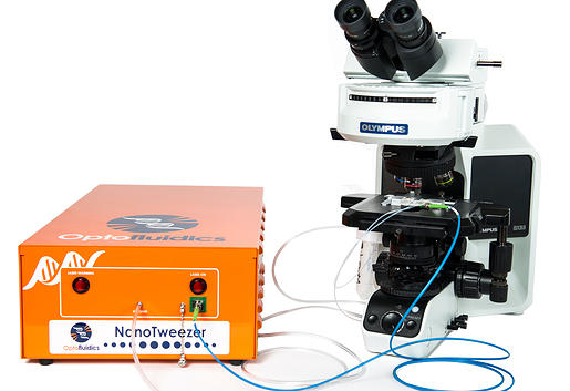
美國Optofluidic品牌nanotweezer微流控納米拉曼光鑷(NanoTweezer Raman)
—光譜分析進行個別納米粒子識別和化學分析(Individual Nanoparticle Identification and Chemical Analysis with Spectroscopy)
進行使用NanoTweezer拉曼光鑷化學分析納米顆粒液體樣品.
微流控技術是在尺度為幾個或上百微米的通道中操縱納升或納升以下流體的技術。
光鑷拉曼光譜技術是將光學囚禁技術與顯微拉曼光譜技術相結合用于細胞或
其它生物大分子、病毒、蛋白的一項新技術本項目將激光光鑷拉曼技術與微流控芯片技術聯(lián)合起來構建了一套全自動化平臺系統(tǒng)。
美國Optofluidic成功的把微流控芯片技術與激光光鑷拉曼技術結合起來,并使流體速度得到了很好的控制,通過圖像識別的方式來判斷細胞是否被俘獲,通過程序來實現(xiàn)俘獲細胞、收集光譜、釋放細胞的完全自動化實驗操作,通過光譜我們可以很方便地區(qū)分正常的血紅細胞與地中海貧血的血紅細胞及其他納米級微粒。
通過圖像識別技術,實現(xiàn)了在微流控芯片的微通道里,由光鑷子將單個活細胞(其他納米粒子)或細胞器長時間固定在焦點附近,進行拉曼光譜分析。用拉曼光鑷子系統(tǒng)實時
監(jiān)測單個活細胞或細胞器的生命過程,能夠在細胞和細胞器水平上,
為研究生命活動的本質提供重要信息
_clip_image002.jpg)
該NanoTweezer拉曼光鑷使研究人員能夠以前所未有的清晰,信號強度和精度來捕獲、可視化和獲取個別納米粒子拉曼光譜信息.
這種創(chuàng)新利用近場光散射法允許NanoTweezer拉曼光譜通過拉曼光譜獲取單個納米粒子的分子指紋。處于消逝場的形式增強信號和最小化背景噪聲,提供了遠遠優(yōu)于比傳統(tǒng)的照明系統(tǒng)的結果。
拉曼信號通常非常弱,難以獲得。拉曼信號通常非常弱,難以獲得。傳統(tǒng)系統(tǒng)使用的傳統(tǒng)激光照射集中在一個塊狀片材料,粉末或溶液使得難以從背景中分離感興趣的材料并準確地表征它。到現(xiàn)在為止的拉曼系統(tǒng)只能被用于分析大量單粒子(幾微米),并在干燥的環(huán)境中。該NanoTweezer拉曼光譜消除這些障礙,使溶液中的單個納米化學識別成為現(xiàn)實。
_clip_image004.jpg)
Perform chemical analysis on a liquid sample of nanoparticles with the NanoTweezer Raman.
納米粒子個體(或小團體)都在使用我們的專利波導的方法暴露于激光,并獲得他們的拉曼光譜。這允許兩種類別分析:
1)未知微粒識別,例如,是否有問題的每個粒子是聚苯乙烯,玻璃,二氧化鈦,等等。
2)一種方法來監(jiān)控用那些經(jīng)過化學改性的藥物顆粒之間的差異,例如,裝載的納米顆粒,觀察降低而引起退化,化學修飾過程的質量控制。
特性列表?
1.對于包括質量控制,環(huán)境分析(納米粒子宿命),藥物遞送,納米毒性藥品和食品制劑研發(fā)的重點領域革命性變革(Revolutionary for key areas of R&D including quality control, environmental analysis (nanoparticle fate), drug delivery, nanotoxicity and drug and food formulations).
2.在其天然溶液環(huán)境中亞微米顆粒識別(Identification of sub-micron particles in their native solution environment).
3.使用小體積(<200μL)微流體流動池直截了當?shù)靥幚順悠?br />
4.用戶不必手動搜索粒子
5.近場光散射技術產(chǎn)生增強信號和最小背景噪音
NanoTweezer使用近場拉曼光譜?
不像其他的拉曼光譜顯微鏡,它只能提供關于微粒或較大的信息,該NanoTweezer可以捕集,可視化,獲得真正納米顆粒拉曼光譜光譜。而不是外部激光聚焦到底物上,它采用近場光泄漏出波導光學地激發(fā)和捕集在其天然環(huán)境中的顆粒。該強光漸逝場的形式導致升高的信號,并比傳統(tǒng)的照明系統(tǒng)背景更少。此外,由于粒子在分析期間被暫時捕獲,一個任意長曝光時間可以獲取。
_clip_image006.jpg)
背景?
Raman Spectroscopy??
Raman Spectroscopy is a powerful analytical?? technique that uses a laser to obtain a chemical?? signature from a material. The interrogating light (a?? single wavelength) interacts with the chemical bonds in?? the material causing an energy shift depending on the?? type of bond. These shifts show up as peaks in a?? Raman spectrum. The spectrum is unique to a?? material (glass, polystyrene, titanium dioxide, etc.) and?? can be considered a fingerprint
Raman Limitations
While Raman Spectroscopy can allow researchers to extract molecular fingerprints, Raman signals are typically very weak and difficult to?
obtain. Traditionally, a laser is focused on a bulk piece of material, powder, or solution making it harder to separate the material of interest?
from the background and characterize it accurately. Until now Raman systems could only be used to look at particles if they are stationary?
(not in solution) and visible (greater than a few microns).?
Near Field Raman Using the NanoTweezer
Unlike other Raman microscopes, which can only provide information about microparticles or larger, the NanoTweezer can trap, visualize,?
and obtain Raman spectra from true nanoparticles. Rather than an external laser focused onto a substrate, it uses near field light leaking?
out of a waveguide to optically excite and trap the particles in their native environments. This is the key breakthrough that enables the?
performance increase. The intense light in the form of an evanescent field leads to heightened signal and less background than traditional?
illumination systems. Additionally, because the particles are temporarily trapped during the analysis, an arbitrarily long exposure time can?
be acquired.
_clip_image008.jpg)
Includes
NanoTweezer with 785 nm laser
Microscope with NIR objectives
Microscope camera
Raman Spectrometer
Raman Software
The Benefits of Near-field Light Scattering
New Insights into Particle Analysis
_clip_image010.jpg)
Nanoparticle behavior and function is inextricably linked to the properties of its surface. Yet until today, no technique has allowed researchers to adequately characterize the properties of the surfaces of their nanoparticles in a detailed, simple and high throughput way. ?Near-field Light Scattering (NLS) enables all three of those by quickly mapping the energy and force landscapes of particle-surface and particle-particle interactions.?
Instead of just generating an overtly simplistic number (like zeta potential or debye length) NLS enables heightened information of these interactions which is key for ensuring proper function and designing stable formulations.?
A quick example to bring this home: properly coated particles allow strong repulsive forces, culminating in more stability and less aggregation.?
NLS is useful for key areas of R&D including drug delivery. In this field, particles need to be designed to be stable as well as perform a key specific function (like bind, block, regulate, etc.) Such design is done at the single particle level, and particles are expected to act on an individual basis rather than an aggregated basis, since aggregated particles either become ineffective or dangerous.?
One key benefit of NLS is that it allows researchers to analyze and distinguish between different aggregation states for the same particle population.
_clip_image012.jpg)
_clip_image014.jpg)
Comparison with State-of-the-Art Methods:
With a myriad of other particle analysis tools available, why use NLS? NLS can be utilized to study a variety of particles and surface phenomena, including charge interactions, uncharged polar, as well as steric interactions. Unlike techniques like Zeta Potential which require a charged surface, NLS works for all types of particle surfaces.? Unlike Zeta potential measurements, which while useful ultimately result in simplistic “collapsed” measurement, NLS measurements reveal magnitude and characteristics of surface interactions at the single particle level through its detailed force and energy maps. Also, existing particle analysis instruments like dynamic light scattering or particle tracking algorithms are useful for predicting accurate particle size, but fail to provide further surface insights. At the same time, existing chemical characterization technologies like UV- VIS and Raman are limited to bulk surfaces or to very large particles in dry environments. NLS works quickly and accurately with nanoparticles in their native suspended states. NLS is the latest form of light scattering, exploiting light localized on a small optical fiber (called a waveguide) to optically excite particles far more efficiently than traditional illumination systems. Briefly, the photons in the evanescent field capture and interact with the selected nanoparticles. 亮點及典型應用:
Some key advantages include:?
1. Universal application
2. Empowering Breakthroughs through Further InsightsHow it works? Well... you see it work!
"The evanescent field can interact with particles much more efficiently than traditional far field systems...we are just starting to see the advantages in near-field light scattering, with nanoparticle surface measurements being a key example," explains Cornell Professor David Erickson.
The particles that interact with the waveguide scatter light so efficiently that one can see them as they approach and propagate along the waveguide due to the unique interactions it allows. Using the ?evanescent field with the latest in nanophotonic design, Optofluidics has made it possible to outperform both manipulation and optical excitation techniques, bringing about the most revolutionary particle surface analysis technique in the market..
美國OptoFluidics顯微鏡納米激光鑷轉換裝置?
_clip_image016.jpg)
*5分鐘將顯微鏡升級為納米級激光光鑷?
三大功能
_clip_image018.jpg) ?
?
_clip_image020.jpg) ?
?
系統(tǒng)技術參數(shù):
1、系統(tǒng)聯(lián)機能力:?
能與科研級正置/倒置顯微鏡聯(lián)用?
能與激光顯微拉曼光譜儀聯(lián)用?
2、捕獲與操縱能力:?
新型納米光鑷系統(tǒng)NanoTweezer,采用世界先進的諧振器波導系統(tǒng),通過微芯片發(fā)出的激光捕獲與操縱納米至微米級的粒子。可以實現(xiàn)多種應用,如操作遠遠小于傳統(tǒng)的光學鑷子的樣品,并保持粒子結構不被破壞;實行新類型的實驗和分析.?
2.1 粒子捕獲操縱尺寸范圍:50nm-5微米?
2.2 操作對象涵蓋了單個細胞、單個分子、病毒、核酸、納米顆粒、碳納米管和蛋白質?
3、激光器:?
3.1激光波長(Wavelength) :1065 nm
3.2激光功率(Optical Power) :0—500 mW連續(xù)可調(diào)?
3.3光纖接口:FC / APC SMPM
3.4光電隔離(Optical Isolation) :33-38 dB
4、顯微鏡流動池:?
4.1最大壓力:20psi (1psi=6.895kPa)
4.2流動速度:80nl/min (min)–1000ml/hr (max)
5、與顯微拉曼光譜儀聯(lián)用附件?
5.1 光鑷與拉曼光譜儀聯(lián)鏈接適配器?
5.2? OD6帶阻濾光片(1064nm)?
6、儀器尺寸:8“(寬)X14”(長)x9“(高)
?