微管相關蛋白1A抗體
產品名稱: 微管相關蛋白1A抗體
英文名稱: MAP1A
產品編號: 1847
產品價格: null
產品產地: 上海
品牌商標: 雅吉
更新時間: null
使用范圍: IHC-P IHC-F Flow-Cyt IF
上海雅吉生物科技有限公司
- 聯系人 :
- 地址 : 上海市閔行區元江路5500號第1幢5658室
- 郵編 :
- 所在區域 : 上海
- 電話 : 158****3937 點擊查看
- 傳真 : 點擊查看
- 郵箱 : yajikit@163.com
?
| 中文名稱 | 微管相關蛋白1A抗體 |
| 別????名 | MAP 1A; MAP-1A; MTAP1A; MAP 1L; MAP1L; MAP1A heavy chain; Map1a; MAP1A_HUMAN; Microtubule associated protein 1 like; Microtubule Associated Protein 1A; Microtubule-associated protein 1A; MTAP1A; Proliferation related protein p80; Proliferation-related protein p80; AI853608; Map1a; moth1; Mtap-1; Mtap1.?? |
| 研究領域 | 腫瘤??細胞生物??免疫學??神經生物學??信號轉導??細胞凋亡??激酶和磷酸酶?? |
| 抗體來源 | Rabbit |
| 克隆類型 | Polyclonal |
| 交叉反應 | Human,?Rat,? (predicted: Mouse,?Chicken,?Dog,?Pig,?Cow,?Horse,?) |
| 產品應用 | IHC-P=1:100-500?IHC-F=1:100-500?Flow-Cyt=1μg /test?IF=1:100-500?(石蠟切片需做抗原修復) not yet tested in other applications. optimal dilutions/concentrations should be determined by the end user. |
| 分?子?量 | 326kDa |
| 細胞定位 | 細胞漿? |
| 性????狀 | Liquid |
| 濃????度 | 1mg/ml |
| 免?疫?原 | KLH conjugated synthetic peptide derived from human MAP1A heavy chain:2651-1750/3014? |
| 亞????型 | IgG |
| 純化方法 | affinity purified by Protein A |
| 儲?存?液 | 0.01M TBS(pH7.4) with 1% BSA, 0.03% Proclin300 and 50% Glycerol. |
| 保存條件 | Shipped at 4℃. Store at -20 °C for one year. Avoid repeated freeze/thaw cycles. |
| PubMed | PubMed |
| 產品介紹 | This gene encodes a protein that belongs to the microtubule-associated protein family. The proteins of this family are thought to be involved in microtubule assembly, which is an essential step in neurogenesis. The product of this gene is a precursor polypeptide that presumably undergoes proteolytic processing to generate the final MAP1A heavy chain and LC2 light chain. Expression of this gene is almost exclusively in the brain. Studies of the rat microtubule-associated protein 1A gene suggested a role in early events of spinal cord development. [provided by RefSeq, Jul 2008] Function: Structural protein involved in the filamentous cross-bridging between microtubules and other skeletal elements. Subunit: 3 different light chains, LC1, LC2 and LC3, can associate with MAP1A and MAP1B proteins. Interacts with TIAM2. Interacts with guanylate kinase-like domain of DLG1, DLG2, DLG4. Binds to CSNK1D. Subcellular Location: Cytoplasm, cytoskeleton (Probable). Tissue Specificity: Brain Post-translational modifications: Phosphorylated by CSNK1D. LC2 is generated from MAP1A by proteolytic processing. Similarity: Belongs to the MAP1 family. SWISS: P78559 Gene ID: 4130 Database links: Entrez Gene: 4130?Human Entrez Gene: 17754?Mouse Entrez Gene: 25152?Rat Omim: 600178?Human SwissProt: P78559?Human SwissProt: Q9QYR6?Mouse SwissProt: P34926?Rat Unigene: 194301?Human Unigene: 619338?Human Unigene: 36501?Mouse Unigene: 11402?Rat Unigene: 41412?Rat Important Note: This product as supplied is intended for research use only, not for use in human, therapeutic or diagnostic applications. ? |
| 產品圖片 | 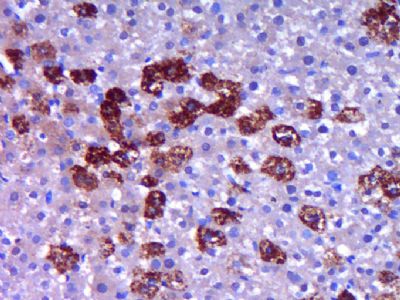 Paraformaldehyde-fixed, paraffin embedded (Rat liver); Antigen retrieval by boiling in sodium citrate buffer (pH6.0) for 15min; Block endogenous peroxidase by 3% hydrogen peroxide for 20 minutes; Blocking buffer (normal goat serum) at 37°C for 30min; Antibody incubation with (MAP1A) Polyclonal Antibody, Unconjugated (bs-1847R) at 1:400 overnight at 4°C, followed by operating according to SP Kit(Rabbit) (sp-0023) instructionsand DAB staining. Paraformaldehyde-fixed, paraffin embedded (Rat liver); Antigen retrieval by boiling in sodium citrate buffer (pH6.0) for 15min; Block endogenous peroxidase by 3% hydrogen peroxide for 20 minutes; Blocking buffer (normal goat serum) at 37°C for 30min; Antibody incubation with (MAP1A) Polyclonal Antibody, Unconjugated (bs-1847R) at 1:400 overnight at 4°C, followed by operating according to SP Kit(Rabbit) (sp-0023) instructionsand DAB staining.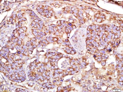 Tissue/cell: human cervical carcinoma; 4% Paraformaldehyde-fixed and paraffin-embedded; Tissue/cell: human cervical carcinoma; 4% Paraformaldehyde-fixed and paraffin-embedded;Antigen retrieval: citrate buffer ( 0.01M, pH 6.0 ), Boiling bathing for 15min; Block endogenous peroxidase by 3% Hydrogen peroxide for 30min; Blocking buffer (normal goat serum,C-0005) at 37℃ for 20 min; Incubation: Anti-MAP1A Polyclonal Antibody, Unconjugated(bs-1847R) 1:500, overnight at 4°C, followed by conjugation to the secondary antibody(SP-0023) and DAB(C-0010) staining 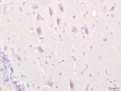 Tissue/cell: rat brain tissue; 4% Paraformaldehyde-fixed and paraffin-embedded; Tissue/cell: rat brain tissue; 4% Paraformaldehyde-fixed and paraffin-embedded;Antigen retrieval: citrate buffer ( 0.01M, pH 6.0 ), Boiling bathing for 15min; Block endogenous peroxidase by 3% Hydrogen peroxide for 30min; Blocking buffer (normal goat serum,C-0005) at 37℃ for 20 min; Incubation: Anti-MAP1A Polyclonal Antibody, Unconjugated(bs-1847R) 1:500, overnight at 4°C, followed by conjugation to the secondary antibody(SP-0023) and DAB(C-0010) staining 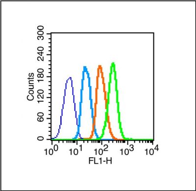 Blank control (blue line): U251 (blue). Blank control (blue line): U251 (blue).Primary Antibody (green line): Rabbit Anti- MAP1A antibody (bs-1847R) Dilution: 1μg /10^6 cells; Isotype Control Antibody (orange line): Rabbit IgG . Secondary Antibody (white blue line): Goat anti-rabbit IgG-PE Dilution: 1μg /test. Protocol The cells were fixed with 2% paraformaldehyde (10 min , then permeabilized) with 90% ice-cold methanol for 20 min on ice. Cells stained with Primary Antibody for 30 min at room temperature. The cells were then incubated in 1 X PBS/2%BSA/10% goat serum to block non-specific protein-protein interactions followed by the antibody for 15 min at room temperature. The secondary antibody used for 40 min at room temperature. Acquisition of 20,000 events was performed. |
?