PARP1 Rabbit Polyclonal Antibody (KO Validated)
產品名稱: PARP1 Rabbit Polyclonal Antibody (KO Validated)
英文名稱: PARP1 Rabbit Polyclonal Antibody (KO Validated)
產品編號: K5264
產品價格: null
產品產地: 上海
品牌商標: 雅吉生物
更新時間: null
使用范圍: WB, IF, ICC
- 聯系人 :
- 地址 : 上海市閔行區元江路5500號第1幢5658室
- 郵編 :
- 所在區域 : 上海
- 電話 : 158****3937 點擊查看
- 傳真 : 點擊查看
- 郵箱 : yajikit@163.com
| 來源 | 用途 | 交叉反應性 | 理論分子量 | 實際分子量 |
| Rabbit | WB, IP, IF, IHC, CHIP | H, M, R | 113kDa | 100/110/125kDa |
WB, Western blot; IP, Immunoprecipitation; IF, Immunofluorescence; IHC, Immunohistochemistry; ICC, Immunocytochemistry; FC, Flow Cytometry; ChIP, Chromatin Immunoprecipitation Assay; ChIP-seq, ChIP-sequencing.
H, Human; M, Mouse; R, Rat; C, Chicken; Cw, Cow; Dg, Dog; Gp, Guinea pig; Hm, Hamster; Hr, Horse; Mk, Monkey; Pg, Pig; Rb, Rabbit; S, Sheep; Z, Zebrafish; All, all species expected.
配套提供了Western一抗稀釋液,可以用于Western檢測或其它適當用途時的一抗稀釋。
建議抗體使用時的稀釋比例如下(實際使用時需根據抗原水平的高低作適當調整):
| WB | IP | IF | IHC | ICC | FC | ChIP | ChIP-seq |
| 1:1000-1:3000 | 1:50-1:200 | 1:50-1:200 | 1:50-1:200 | 1:50-1:200 | - | 1:50-1:200 | - |
抗體詳細信息如下::
| About this Antibody | |
| Name | PARP1 Rabbit Polyclonal Antibody (KO Validated) |
| Category | Rabbit Polyclonal Antibody (pAb); Primary antibody |
| Isotype | IgG |
| Purification method | Affinity purification |
| Positive samples | Jurkat, HeLa, RAW264.7, C6, Mouse testis, Mouse brain |
| Cellular location | Nucleus, nucleolus |
| Customer validation | WB (Human) |
| About the Immunogen | |
| Immunogen | Recombinant fusion protein of human PARP1 (NP_001609.2). |
| Sequence | DQQKVKKTAEAGGVTGKGQDGIGSKAEKTLGDFAAEYAKSNRSTCKGCMEKIEKGQVRLSKKMVDPEKPQLGMIDRWYHPGCFVKNREELGFRPEYSASQLKGFSLLATEDKEALKKQLPGVKSEGKRKGDEVDGVDEVAKKKSKKEKDKDSKLEKALKAQNDLIWNIKDELKKVCSTNDLKELLIFNKQQVPSGESAILDRVADGMVFGALLPCEECSGQLVFKSDAYYCTGDVTAWTKCMVKTQTPNRKEWVTPKEFREISYLKKLKVKKQDRIFPPETSASVAATPPPSTASAPAAVNSSASADKPL |
| Gene ID | 142 |
| Swiss Prot | P09874 |
| Synonyms | PARP1; ADPRT; ADPRT 1; ADPRT1; ARTD1; PARP; PARP-1; PPOL; pADPRT-1; poly(ADP-ribose) polymerase 1 |
| Category | Death Receptor Signaling; NF-κB Signaling |
| Background | PARP1, a 116 kDa nuclear poly (ADP-ribose) polymerase, appears to be involved in DNA repair in response to environmental stress. This protein can be cleaved by many ICE-like caspases in vitro and is one of the main cleavage targets of caspase-3 in vivo . In human PARP, the cleavage occurs between Asp214 and Gly215, which separates the PARP amino-terminal DNA binding domain (24 kDa) from the carboxy-terminal catalytic domain (89 kDa). PARP helps cells to maintain their viability; cleavage of PARP facilitates cellular disassembly and serves as a marker of cells undergoing apoptosis. |
包裝清單:
| 產品編號 | 產品名稱 | 包裝 |
| AF5264 | PARP1 Rabbit Polyclonal Antibody (KO Validated) | 50μl |
| AZ050 | Western一抗稀釋液 | 50ml |
| — | 說明書 | 1份 |
保存條件:
PARP1 Rabbit Polyclonal Antibody (KO Validated) -20oC保存,Western一抗稀釋液-20oC或4oC保存,一年有效。Western一抗稀釋液優先推薦4oC保存,長期不使用可以考慮-20oC保存,但凍融可能會導致出現輕微的渾濁和少量不溶物。
注意事項:
如果本抗體用于Western blot (WB)、免疫熒光(IF)、免疫細胞化學(ICC)等實驗,請注意回收使用過的稀釋抗體。回收的抗體通常至少可以重復使用5-10次。稀釋后的抗體,包括已經使用過的稀釋抗體,請4℃保存。
回收后重復使用的抗體,使用方法同新鮮稀釋的抗體。如果在重復使用過程中發現抗體出現輕微混濁現象,可以10,000g離心1-3分鐘,取上清用于后續檢測。如果回收的抗體出現明顯的絮狀物或長霉長菌等情況,則可以考慮廢棄該抗體。
提供的Western一抗稀釋液也可以用于免疫熒光(IF)、免疫組化(IHC)、免疫細胞化學(ICC)等適當用途。如果希望獲得最佳的檢測效果,請考慮使用上述檢測專用的一抗稀釋液。
本產品僅限于專業人員的科學研究用,不得用于臨床診斷或治療,不得用于食品或藥品,不得存放于普通住宅內。
為了您的安全和健康,請穿實驗服并戴一次性手套操作。
代表性圖片:
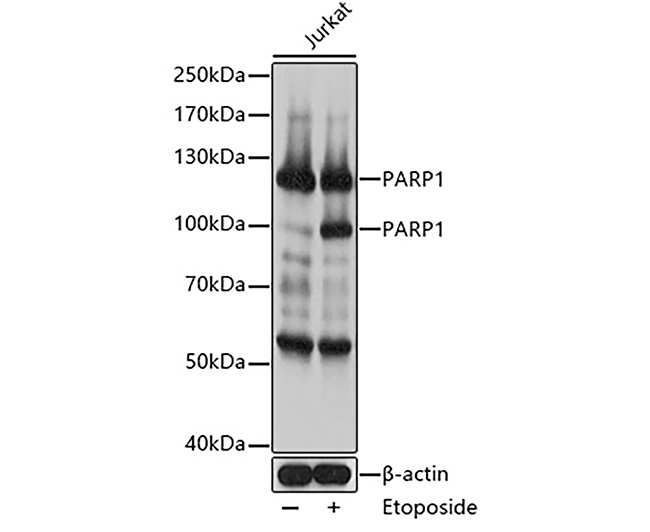 | 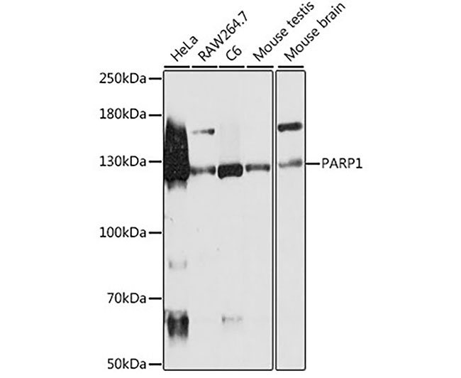 |
| Fig. 1. Western blot analysis of extracts of various cell lines, using PARP1 antibody at 1:500 dilution. Secondary antibody: HRP-labeled Goat Anti-Rabbit IgG(H+L) (A0208) at 1:1000 dilution. Lysates/proteins: 25μg per lane. Blocking buffer: QuickBlock? Blocking Buffer (P0231). Detection: BeyoECL Star (P0018A). Exposure time:1s. | Fig. 2. Western blot analysis of extracts of Jurkat cells, using PARP1 antibody at 1:1000 dilution. Secondary antibody: HRP-labeled Goat Anti-Rabbit IgG(H+L) (A0208) at 1:1000 dilution. Lysates/proteins: 25μg per lane. Blocking buffer: QuickBlock? Blocking Buffer (P0231). Detection: BeyoECL Star (P0018A). Exposure time:1s. |
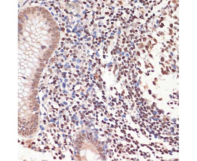 | 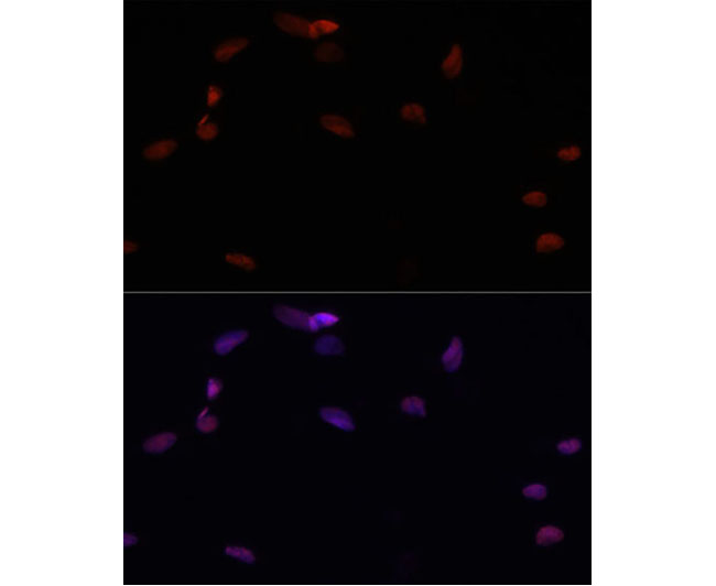 |
| Fig. 3. Immunohistochemistry of paraffin-embedded human appendix using PARP1 antibody at dilution of 1:200 (40x lens). | Fig. 4. Confocal immunofluorescence analysis of U-2 OS cells using PARP1 Polyclonal Antibody at dilution of 1:200. Blue: DAPI for nuclear staining. |