半胱氨酸蛋白酶抗體
產(chǎn)品名稱(chēng): 半胱氨酸蛋白酶抗體
英文名稱(chēng): HSD17B3
產(chǎn)品編號(hào): 3905
產(chǎn)品價(jià)格: null
產(chǎn)品產(chǎn)地: 上海
品牌商標(biāo): 雅吉
更新時(shí)間: null
使用范圍: WB ELISA IHC-P Flow-Cyt
上海雅吉生物科技有限公司
- 聯(lián)系人 :
- 地址 : 上海市閔行區(qū)元江路5500號(hào)第1幢5658室
- 郵編 :
- 所在區(qū)域 : 上海
- 電話(huà) : 158****3937 點(diǎn)擊查看
- 傳真 : 點(diǎn)擊查看
- 郵箱 : yajikit@163.com
?
| 中文名稱(chēng) | 半胱氨酸蛋白酶抗體 |
| 別????名 | AEP; Asparaginyl endopeptidase; EC 3.4.22.34; LGMN; LGMN1; Protease, cysteine 1; protease, cysteine, 1 (legumain); PRSC1; LGMN_HUMAN.?? |
| 研究領(lǐng)域 | 腫瘤??細(xì)胞生物??免疫學(xué)??細(xì)胞凋亡??激酶和磷酸酶?? |
| 抗體來(lái)源 | Rabbit |
| 克隆類(lèi)型 | Polyclonal |
| 交叉反應(yīng) | Human,?Mouse,?Rat,? (predicted: Dog,?Cow,?Horse,?) |
| 產(chǎn)品應(yīng)用 | WB=1:500-2000?ELISA=1:500-1000?IHC-P=1:100-500?Flow-Cyt=3ug/Test?(石蠟切片需做抗原修復(fù)) not yet tested in other applications. optimal dilutions/concentrations should be determined by the end user. |
| 分?子?量 | 46kDa |
| 細(xì)胞定位 | 細(xì)胞漿? |
| 性????狀 | Liquid |
| 濃????度 | 1mg/ml |
| 免?疫?原 | KLH conjugated synthetic peptide derived from human Legumain:201-300/433? |
| 亞????型 | IgG |
| 純化方法 | affinity purified by Protein A |
| 儲(chǔ)?存?液 | 0.01M TBS(pH7.4) with 1% BSA, 0.03% Proclin300 and 50% Glycerol. |
| 保存條件 | Shipped at 4℃. Store at -20 °C for one year. Avoid repeated freeze/thaw cycles. |
| PubMed | PubMed |
| 產(chǎn)品介紹 | This gene encodes a cysteine protease that has a strict specificity for hydrolysis of asparaginyl bonds. This enzyme may be involved in the processing of bacterial peptides and endogenous proteins for MHC class II presentation in the lysosomal/endosomal systems. Enzyme activation is triggered by acidic pH and appears to be autocatalytic. Protein expression occurs after monocytes differentiate into dendritic cells. A fully mature, active enzyme is produced following lipopolysaccharide expression in mature dendritic cells. Overexpression of this gene may be associated with the majority of solid tumor types. This gene has a pseudogene on chromosome 13. Several alternatively spliced transcript variants have been described, but the biological validity of only two has been determined. These two variants encode the same isoform. [provided by RefSeq, Jul 2008]. Function: Has a strict specificity for hydrolysis of asparaginyl bonds. Can also cleave aspartyl bonds slowly, especially under acidic conditions. May be involved in the processing of proteins for MHC class II antigen presentation in the lysosomal/endosomal system. Subcellular Location: Lysosome (By similarity). Tissue Specificity: Ubiquitous. Particularly abundant in kidney, heart and placenta. Post-translational modifications: Glycosylated. Similarity: Belongs to the peptidase C13 family. SWISS: Q99538 Gene ID: 5641 Database links: Entrez Gene: 5641?Human Entrez Gene: 19141?Mouse Entrez Gene: 63865?Rat Omim: 602620?Human SwissProt: Q99538?Human SwissProt: O89017?Mouse SwissProt: Q9R0J8?Rat Unigene: 726036?Human Unigene: 17185?Mouse Unigene: 206021?Rat Important Note: This product as supplied is intended for research use only, not for use in human, therapeutic or diagnostic applications. ? |
| 產(chǎn)品圖片 | 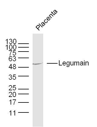 Sample: Placenta (Mouse) Lysate at 40 ug Sample: Placenta (Mouse) Lysate at 40 ugPrimary: Anti-Legumain (bs-3907R) at 1/300 dilution Secondary: IRDye800CW Goat Anti-Rabbit IgG at 1/20000 dilution Predicted band size: 46 kD Observed band size: 55 kD 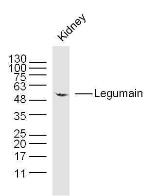 Sample: Kidney (Mouse) Lysate at 40 ug Sample: Kidney (Mouse) Lysate at 40 ugPrimary: Anti- Legumain (bs-3907R) at 1/300 dilution Secondary: IRDye800CW Goat Anti-Rabbit IgG at 1/20000 dilution Predicted band size: 46 kD Observed band size: 55 kD 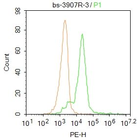 Blank control: HL60. Primary Antibody (green line): Rabbit Anti-Legumain antibody (bs-3907R) Dilution: 3μg /10^6 cells; Isotype Control Antibody (orange line): Rabbit IgG . Secondary Antibody : Goat anti-rabbit IgG-PE Dilution: 1μg /test. Protocol The cells were fixed with 4% PFA (10min at room temperature)and then permeabilized with PBST for 20 min at room temperature. The cells were then incubated in 5%BSA to block non-specific protein-protein interactions for 30 min at at room temperature .Cells stained with Primary Antibody for 30 min at room temperature. The secondary antibody used for 40 min at room temperature. Acquisition of 20,000 events was performed. Blank control: HL60. Primary Antibody (green line): Rabbit Anti-Legumain antibody (bs-3907R) Dilution: 3μg /10^6 cells; Isotype Control Antibody (orange line): Rabbit IgG . Secondary Antibody : Goat anti-rabbit IgG-PE Dilution: 1μg /test. Protocol The cells were fixed with 4% PFA (10min at room temperature)and then permeabilized with PBST for 20 min at room temperature. The cells were then incubated in 5%BSA to block non-specific protein-protein interactions for 30 min at at room temperature .Cells stained with Primary Antibody for 30 min at room temperature. The secondary antibody used for 40 min at room temperature. Acquisition of 20,000 events was performed.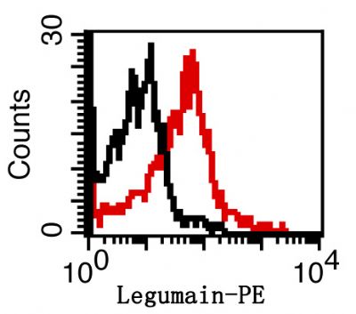 Overlay histogram showing Mouse spleen cells stained with bs-3907-PE (red line). The cells were fixed with 1% paraformaldehyde (10 min).The cells were then incubated with the antibody (bs-3907R-PE, 2ug/1x106cells) for 30 min at 22-25℃. Isotype control antibody (black line) was rabbit IgG (2ug/1x106cells) used under the same conditions. Acquisition of events were used for analysis. Overlay histogram showing Mouse spleen cells stained with bs-3907-PE (red line). The cells were fixed with 1% paraformaldehyde (10 min).The cells were then incubated with the antibody (bs-3907R-PE, 2ug/1x106cells) for 30 min at 22-25℃. Isotype control antibody (black line) was rabbit IgG (2ug/1x106cells) used under the same conditions. Acquisition of events were used for analysis. |
| ? | ? |
| ? | ? |
| ? | ? |
| ? | ? |
| ? | ? |
| ? | ? |
| ? | ? |
| ? | ? |
| ? | ? |
| ? | ? |
| ? | ? |
| ? | ? |
| ? | ? |
| ? | ? |
| ? | ? |
| ? | ? |
| ? | ? |