Phospho-CHEK1 (Ser317) Rabbit Polyclonal Antibody
產品名稱: Phospho-CHEK1 (Ser317) Rabbit Polyclonal Antibody
英文名稱: Phospho-CHEK1 (Ser317) Rabbit Polyclonal Antibody
產品編號: K5770
產品價格: null
產品產地: 上海
品牌商標: 雅吉生物
更新時間: null
使用范圍: WB, IF, ICC
- 聯系人 :
- 地址 : 上海市閔行區元江路5500號第1幢5658室
- 郵編 :
- 所在區域 : 上海
- 電話 : 158****3937 點擊查看
- 傳真 : 點擊查看
- 郵箱 : yajikit@163.com
| 來源 | 用途 | 交叉反應性 | 理論分子量 | 實際分子量 |
| Rabbit | WB, IF | H | 43/50/54kDa | 60kDa |
WB, Western blot; IP, Immunoprecipitation; IF, Immunofluorescence; IHC, Immunohistochemistry; ICC, Immunocytochemistry; FC, Flow Cytometry; ChIP, Chromatin Immunoprecipitation Assay; ChIP-seq, ChIP-sequencing.
H, Human; M, Mouse; R, Rat; C, Chicken; Cw, Cow; Dg, Dog; Gp, Guinea pig; Hm, Hamster; Hr, Horse; Mk, Monkey; Pg, Pig; Rb, Rabbit; S, Sheep; Z, Zebrafish; All, all species expected.
配套提供了Western一抗稀釋液,可以用于Western檢測或其它適當用途時的一抗稀釋。
建議抗體使用時的稀釋比例如下(實際使用時需根據抗原水平的高低作適當調整):
| WB | IP | IF | IHC | ICC | FC | ChIP | ChIP-seq |
| 1:500-1:2000 | - | 1:50-1:200 | - | - | - | - | - |
抗體詳細信息如下::
| About this Antibody | |
| Name | Phospho-CHEK1 (Ser317) Rabbit Polyclonal Antibody |
| Category | Rabbit Polyclonal Antibody (pAb); Primary antibody |
| Isotype | IgG |
| Purification method | Affinity purification |
| Positive samples | MCF7 |
| Cellular location | Cytoplasm, Nucleus, centrosome, cytoskeleton, microtubule organizing center |
| Customer validation | - |
| About the Immunogen | |
| Immunogen | A synthetic phosphorylated peptide around S317 of human CHEK1 (NP_001265.2). |
| Sequence | SSSQP |
| Gene ID | 1111 |
| Swiss Prot | O14757 |
| Synonyms | CHEK1; CHK1; checkpoint kinase 1 |
| Category | Regulation of Apoptosis; G1/S Checkpoint; G2M/DNA Damage Checkpoint |
| Background | Chk1/ CHEK1 kinase acts downstream of ATM/ATR kinase and plays an important role in DNA damage checkpoint control, embryonic development, and tumor suppression. Activation of Chk1 involves phosphorylation at Ser317 and Ser345 by ATM/ATR, followed by autophosphorylation of Ser296. Activation occurs in response to blocked DNA replication and certain forms of genotoxic stress. While phosphorylation at Ser345 serves to localize Chk1 to the nucleus following checkpoint activation, phosphorylation at Ser317 along with site-specific phosphorylation of PTEN allows for re-entry into the cell cycle following stalled DNA replication. Chk1 exerts its checkpoint mechanism on the cell cycle, in part, by regulating the cdc25 family of phosphatases. Chk1 phosphorylation of cdc25A targets it for proteolysis and inhibits its activity through 14-3-3 binding. Activated Chk1 can inactivate cdc25C via phosphorylation at Ser216, blocking the activation of cdc2 and transition into mitosis. Centrosomal Chk1 has been shown to phosphorylate cdc25B and inhibit its activation of CDK1-cyclin B1, thereby abrogating mitotic spindle formation and chromatin condensation. Furthermore, Chk1 plays a role in spindle checkpoint function through regulation of aurora B and BubR1. Research studies have implicated Chk1 as a drug target for cancer therapy as its inhibition leads to cell death in many cancer cell lines. |
包裝清單:
| 產品編號 | 產品名稱 | 包裝 |
| AF5770 | Phospho-CHEK1 (Ser317) Rabbit Polyclonal Antibody | 50μl |
| AZ050 | Western一抗稀釋液 | 50ml |
| — | 說明書 | 1份 |
保存條件:
Phospho-CHEK1 (Ser317) Rabbit Polyclonal Antibody -20oC保存,Western一抗稀釋液-20oC或4oC保存,一年有效。Western一抗稀釋液優先推薦4oC保存,長期不使用可以考慮-20oC保存,但凍融可能會導致出現輕微的渾濁和少量不溶物。
注意事項:
如果本抗體用于Western blot (WB)、免疫熒光(IF)、免疫細胞化學(ICC)等實驗,請注意回收使用過的稀釋抗體。回收的抗體通常至少可以重復使用5-10次。稀釋后的抗體,包括已經使用過的稀釋抗體,請4℃保存。
回收后重復使用的抗體,使用方法同新鮮稀釋的抗體。如果在重復使用過程中發現抗體出現輕微混濁現象,可以10,000g離心1-3分鐘,取上清用于后續檢測。如果回收的抗體出現明顯的絮狀物或長霉長菌等情況,則可以考慮廢棄該抗體。
提供的Western一抗稀釋液也可以用于免疫熒光(IF)、免疫組化(IHC)、免疫細胞化學(ICC)等適當用途。如果希望獲得最佳的檢測效果,請考慮使用上述檢測專用的一抗稀釋液。
本產品僅限于專業人員的科學研究用,不得用于臨床診斷或治療,不得用于食品或藥品,不得存放于普通住宅內。
為了您的安全和健康,請穿實驗服并戴一次性手套操作。
代表性圖片:
 | 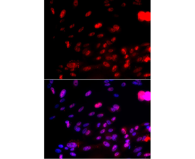 |
| Fig. 1. Western blot analysis of extracts of MCF7 cell line, using Phospho-CHEK1-S317 antibody at 1:10000 dilution. Lysates/proteins: 25μg per lane. Blocking buffer: QuickBlock? Blocking Buffer (P0231). | Fig. 2. Immunofluorescence analysis of MCF-7 cells using Phospho-CHEK1-S317 antibody. Blue: DAPI for nuclear staining. |
 | |
| Fig. 3. Immunofluorescence analysis of U2OS cells using Phospho-CHEK1-S317 antibody. Blue: DAPI for nuclear staining. | |