小膠質(zhì)細(xì)胞(microglia)是中樞神經(jīng)系統(tǒng)的常駐免疫細(xì)胞,占腦內(nèi)膠質(zhì)細(xì)胞總數(shù)的5%–20%,自被神經(jīng)科學(xué)家首次描述以來,其核心功能逐漸被解鎖。它可持續(xù)伸展突起監(jiān)視微環(huán)境,是腦內(nèi)穩(wěn)態(tài)維持、損傷修復(fù)、神經(jīng)發(fā)育的關(guān)鍵角色[1]。
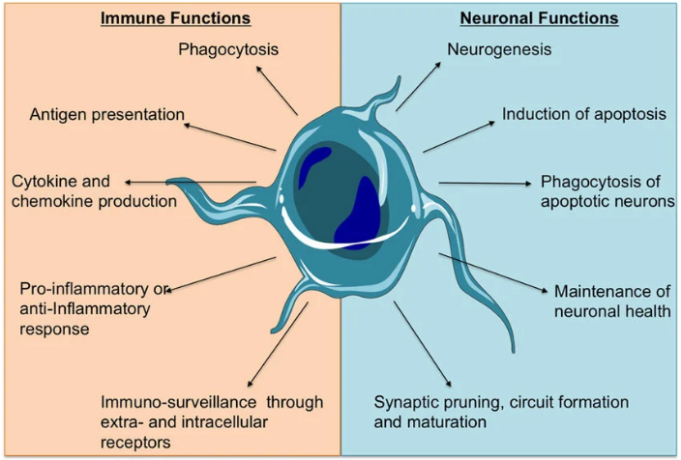
小膠質(zhì)細(xì)胞功能描述[2]
生理狀態(tài)下,小膠質(zhì)細(xì)胞負(fù)責(zé)清除凋亡神經(jīng)元、錯誤折疊蛋白和細(xì)胞碎片,參與突觸修剪以塑造神經(jīng)環(huán)路;病理狀態(tài)下,它會快速響應(yīng)炎癥、感染或損傷,通過分泌細(xì)胞因子、趨化因子等調(diào)控免疫反應(yīng),既可以發(fā)揮神經(jīng)保護(hù)作用,過度活化也可能誘發(fā)神經(jīng)炎癥,推動阿爾茨海默病(AD)、帕金森病(PD)等疾病進(jìn)展[1]。
此前,小膠質(zhì)細(xì)胞研究多依賴原代培養(yǎng),但原代細(xì)胞來源稀缺、體外培養(yǎng)難度大,且難以模擬人體病理狀態(tài)下的細(xì)胞表型,限制了神經(jīng)科研的推進(jìn)[3]。而iPSC技術(shù)的興起,為小膠質(zhì)細(xì)胞的體外標(biāo)準(zhǔn)化培養(yǎng)提供了全新解決方案,iPSC誘導(dǎo)小膠質(zhì)細(xì)胞(iPSC-MG)的研究也逐漸深入并向臨床靠近。
iPSC-MG研究進(jìn)展
在技術(shù)優(yōu)化與模型構(gòu)建上,Stanton KC等首次將iPSC來源的小膠質(zhì)細(xì)胞與神經(jīng)元、星形膠質(zhì)細(xì)胞等六類細(xì)胞,整合到3D水凝膠構(gòu)建“迷你人腦”模型,具備成熟血腦屏障和免疫功能,解決傳統(tǒng)類器官細(xì)胞類型不全、缺氧壞死的痛點(diǎn),是目前最貼近真實(shí)人腦微環(huán)境的體外模型,可用于神經(jīng)疾病機(jī)制的精準(zhǔn)研究[4]。
Liu S, et al篩選出6種轉(zhuǎn)錄因子組合,無需feeder細(xì)胞,4天內(nèi)即可將人iPSC高效誘導(dǎo)為功能成熟的小膠質(zhì)細(xì)胞,大幅縮短誘導(dǎo)周期(傳統(tǒng)方案2-4周),轉(zhuǎn)錄組特征與原代小膠質(zhì)細(xì)胞高度匹配,顯著提升誘導(dǎo)標(biāo)準(zhǔn)化程度,適合高通量實(shí)驗[5]。
在應(yīng)用與轉(zhuǎn)化研究上,Chadarevian et al等人通過基因編輯改造iPSC誘導(dǎo)的小膠質(zhì)細(xì)胞,實(shí)現(xiàn)治療性蛋白在中樞神經(jīng)系統(tǒng)的廣泛遞送,同時可用于AD等神經(jīng)退行性疾病的藥物篩選和細(xì)胞治療探索,為臨床轉(zhuǎn)化提供關(guān)鍵依據(jù)[6]。
斯坦福大學(xué)團(tuán)隊開發(fā)無需骨髓消融的iPSC來源小膠質(zhì)細(xì)胞替換療法,在Sandhoff病(溶酶體貯積癥)小鼠模型中驗證有效,將小鼠壽命延長近一倍,證實(shí)iPSC誘導(dǎo)小膠質(zhì)細(xì)胞在神經(jīng)疾病細(xì)胞治療中的可行性,避免傳統(tǒng)骨髓移植的嚴(yán)重副作用,為“現(xiàn)貨型”腦疾病療法提供新方向[7]。
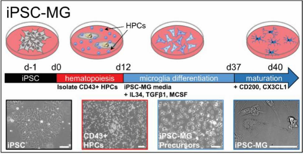
iPSC誘導(dǎo)小膠質(zhì)細(xì)胞示意圖[8]
iPSC-MG核心調(diào)控因子
IL-34(白細(xì)胞介素-34):核心誘導(dǎo)與成熟
與CSF-1R(M-CSFR)結(jié)合,是小膠質(zhì)細(xì)胞分化、存活與功能成熟的關(guān)鍵因子,能增強(qiáng)小膠質(zhì)細(xì)胞的吞噬功能和炎癥響應(yīng)能力[5]。在iPSC誘導(dǎo)體系中,IL-34可促進(jìn)造血祖細(xì)胞向小膠質(zhì)細(xì)胞譜系分化,維持成熟小膠質(zhì)細(xì)胞的表型(如Iba1、CD11b陽性)[9][10]。
M-CSF(巨噬細(xì)胞集落刺激因子):協(xié)同誘導(dǎo)
與IL-34共享CSF-1R受體,協(xié)同IL-34調(diào)控iPSC向小膠質(zhì)細(xì)胞分化,促進(jìn)造血祖細(xì)胞增殖,為小膠質(zhì)細(xì)胞的分化提供基礎(chǔ)[10]。
TGF-β1(轉(zhuǎn)化生長因子β1):表型維持
調(diào)控小膠質(zhì)細(xì)胞的表型成熟,抑制其向巨噬細(xì)胞樣表型轉(zhuǎn)化[10]。在iPSC誘導(dǎo)體系中,TGF-β1可增強(qiáng)小膠質(zhì)細(xì)胞突觸修剪和吞噬功能,確保誘導(dǎo)出的小膠質(zhì)細(xì)胞更貼近體內(nèi)生理狀態(tài)[9][10]。
CD200&CX3CL1:協(xié)同誘導(dǎo),靜息調(diào)控
CD200通過與小膠質(zhì)細(xì)胞表面CD200R1結(jié)合,抑制過度炎癥活化、促進(jìn)細(xì)胞成熟,維持iPSC來源小膠質(zhì)細(xì)胞的生理表型,同時提升細(xì)胞吞噬功能和突觸修剪能力,解決誘導(dǎo)過程中小膠質(zhì)細(xì)胞活化異常、功能紊亂的痛點(diǎn),適配神經(jīng)退行性疾病模型構(gòu)建[11]。
CX3CL1通過與小膠質(zhì)細(xì)胞表面CX3CR1特異性結(jié)合,雙向調(diào)控細(xì)胞分化與功能——誘導(dǎo)階段可促進(jìn)iPSC來源造血祖細(xì)胞向小膠質(zhì)細(xì)胞譜系定向分化;成熟階段可調(diào)控細(xì)胞遷移能力,維持小膠質(zhì)細(xì)胞的靜息態(tài)表型,抑制過度炎癥活化,同時增強(qiáng)其吞噬Aβ蛋白和突觸修剪的功能,適配神經(jīng)退行性疾病和神經(jīng)炎癥模型構(gòu)建[12]。
近岸蛋白可提供iPSC誘導(dǎo)小膠質(zhì)細(xì)胞過程中用到的一系列細(xì)胞因子,包括IL-34、TGF-β1、M-CSF、CD200、CX3CL1等,以及iPSC擴(kuò)增培養(yǎng)常用Vitronectin、Laminin521、Laminin511等基質(zhì)蛋白,高活性,高穩(wěn)定性,高批間一致性,助力高效穩(wěn)定的小膠質(zhì)細(xì)胞研究!
小膠質(zhì)細(xì)胞培養(yǎng)—
近岸蛋白細(xì)胞因子引用文獻(xiàn)(部分):
-
Coburn, Morgan A et al. “Human microglia differentially respond to β-amyloid, tau, and combined Alzheimer's disease pathologies in vivo.” Alzheimer's & dementia : the journal of the Alzheimer's Association vol. 21,11 (2025): e70930.CD200(Cat. No.:C311,100 ng/mL).
-
Lish, Alexandra M et al. “Astrocyte induction of disease-associated microglia is suppressed by acute exposure to fAD neurons in human iPSC triple cultures.” Cell reports vol. 44,6 (2025): 115777. CD200(Cat. No.:C311,100 ng/mL).
-
Iyer, Abhirami K et al. “Cell autonomous microglia defects in a stem cell model of frontotemporal dementia tau.” Molecular psychiatry vol. 30,10 (2025): 4776-4791.CD200(Cat. No.:C311,100 ng/mL).
-
Gao, Mei-Ling et al. “Retinal Organoid Microenvironment Enhanced Bioactivities of Microglia-Like Cells Derived From HiPSCs.” Investigative ophthalmology & visual science vol. 65,12 (2024): 19.IL-3(Cat. No.:CX90,25 ng/mL ),M-CSF(Cat. No.:C417,25 ng/mL)
-
Guan, Xin et al. “Microglial CMPK2 promotes neuroinflammation and brain injury after ischemic stroke.” Cell reports. Medicine vol. 5,5 (2024): 101522. doi:10.1016/j.xcrm.2024.101522.M-CSF(Cat. No.:CB34).
-
Wang, Zhijie et al. “Clock Gene Nr1d1 Alleviates Retinal Inflammation Through Repression of Hmga2 in Microglia.” Journal of inflammation research vol. 14 5901-5918. 11 Nov. 2021, doi:10.2147/JIR.S326091.M-CSF(Cat. No.:CB34,10 ng/mL).
-
Yu, Shui-Sheng et al. “M1-type microglia can induce astrocytes to deposit chondroitin sulfate proteoglycan after spinal cord injury.” Neural regeneration research vol. 17,5 (2022): 1072-1079. doi:10.4103/1673-5374.324858.TGF-β1(Cat. No.:CK33,100 pg/mL).
數(shù)據(jù)展示
Recombinant Human IL-34 (Low Endotoxin,0.01EU/ug)
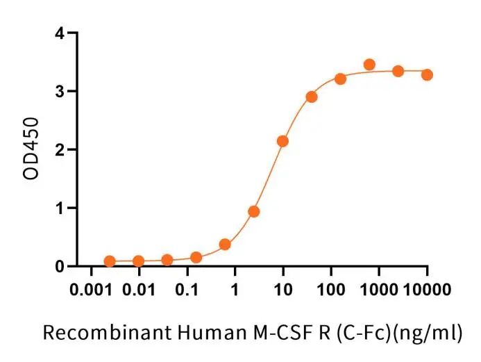
Recombinant Human IL-34 (C-6His, Low Endotoxin)(Cat.No.:C51R) at 2 μg/ml (100 μl/well) can bind Recombinant Human M-CSF R (C-Fc)(Cat.No.:CS42).The ED50 of Recombinant Human M-CSF R (C-Fc)(Cat.No.:CS42) is 6.22 ng/ml.
Recombinant Human TGF-beta 1(引用文獻(xiàn)147篇)
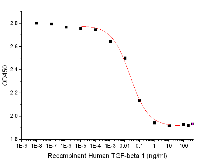
Measured by its ability to inhibit the IL-4-dependent proliferation of TF?1 human erythroleukemic cells. The ED50 for this effect is 4-40 pg/ml.
Recombinant Human M-CSF (引用文獻(xiàn)32篇)
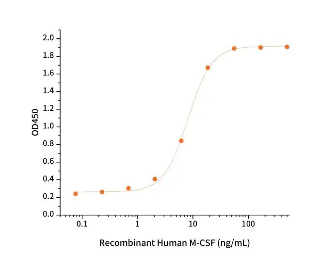
Measured in a cell proliferation assay using M-NFS-60 mouse lymphoblast cells.The ED50 for this effect is 2-10 ng/mL.
小膠質(zhì)細(xì)胞培養(yǎng)相關(guān)產(chǎn)品
|
目錄號 |
產(chǎn)品名稱 |
|
C51R |
Recombinant Human IL-34 |
|
C417 |
Recombinant Human M-CSF |
|
CB34 |
Recombinant Mouse M-CSF |
|
C311 |
Recombinant Human CD200 |
|
CA59 |
Recombinant Human TGF-beta 1 |
|
CK33 |
Recombinant Mouse/Rat TGF-beta 1 |
|
C461 |
Recombinant Human CX3CL1 |
|
CI38 |
Recombinant Mouse CX3CL1 |
參考文獻(xiàn)
[1]Brüll M, et al. Differential Responses of Human iPSC-Derived Microglia to Stimulation with Diverse Inflammogens. Cells. 2025;14(21):1687. DOI: 10.3390/cells14211687.
[2]Silvin, Aymeric, and Florent Ginhoux. “Microglia heterogeneity along a spatio-temporal axis: More questions than answers.” Glia vol. 66,10 (2018): 2045-2057. doi:10.1002/glia.23458.
[3]Hasselmann J, et al. Human iPSC-derived microglia: A growing toolset to study the brain’s innate immune cells. Glia. 2020;68(4):721–739. PMID: 31926038; DOI: 10.1002/glia.23781.
[4]Stanton KC, Sano T, Ma M, et al. Engineered 3D immuno-glial-neurovascular human miBrain model. Proceedings of the National Academy of Sciences of the United States of America (PNAS), 2025, 122(4): e2311455122. DOI: 10.1073/pnas.2311455122.
[5]Liu, Songlei et al. “Iterative transcription factor screening enables rapid generation of microglia-like cells from human iPSC.” Nature communications vol. 16,1 5136. 10 Jun. 2025, doi:10.1038/s41467-025-59596-3.
[6]Chadarevian et al. Harnessing human iPSC-microglia for CNS-wide delivery of disease-modifying proteins. Cell Stem Cell. 2025;32(6):914–934. DOI: 10.1016/j.stem.2025.04.008.
[7]Mader, Marius Marc-Daniel et al. “Therapeutic genetic restoration through allogeneic brain microglia replacement.” Nature vol. 646,8086 (2025): 903-912. doi:10.1038/s41586-025-09461-6.
[8]Rai, Mohammad A et al. “Comparative analysis of human microglial models for studies of HIV replication and pathogenesis.” Retrovirology vol. 17,1 35. 19 Nov. 2020, doi:10.1186/s12977-020-00544-y.
[9]Lehoux M, et al. The Generation and Functional Characterization of Human Microglia-Like Cells Derived from iPS and Embryonic Stem Cells. Methods Mol Biol. 2023;PMID: 37300767; DOI: 10.1007/978-1-0716-3287-1_6.
[10] Hasselmann, Jonathan, and Mathew Blurton-Jones. “Human iPSC-derived microglia: A growing toolset to study the brain's innate immune cells.” Glia vol. 68,4 (2020): 721-739. doi:10.1002/glia.23781.
[11]Song J, et al. CD200 regulates the maturation and functional homeostasis of iPSC-derived microglia via CD200R1 signaling. Stem Cell Research & Therapy. 2024;15(1):128. DOI: 10.1186/s13287-024-03521-9.
[12]Wang L, et al. CX3CL1 supplementation improves the functional fidelity of iPSC-derived microglia in a 3D brain organoid model. Stem Cells and Development. 2023;32(15):987–1000. PMID: 37472892; DOI: 10.1089/scd.2023.0052.