
細胞表面的PS外翻在1995年首次報道為細胞凋亡的顯著特征,并被大量實驗引用,長期以來,PS的外翻被認為是細胞凋亡中不可逆的行為,然而近年來文獻報道PS的外翻是可逆的,不僅存在于凋亡細胞中,也存在于其他細胞形態中。pSIVA-IANBD技術的出現可以實時追蹤PS的外翻,使科研工作者獲得更多不同類型細胞死亡中的細胞膜極性變化信息。
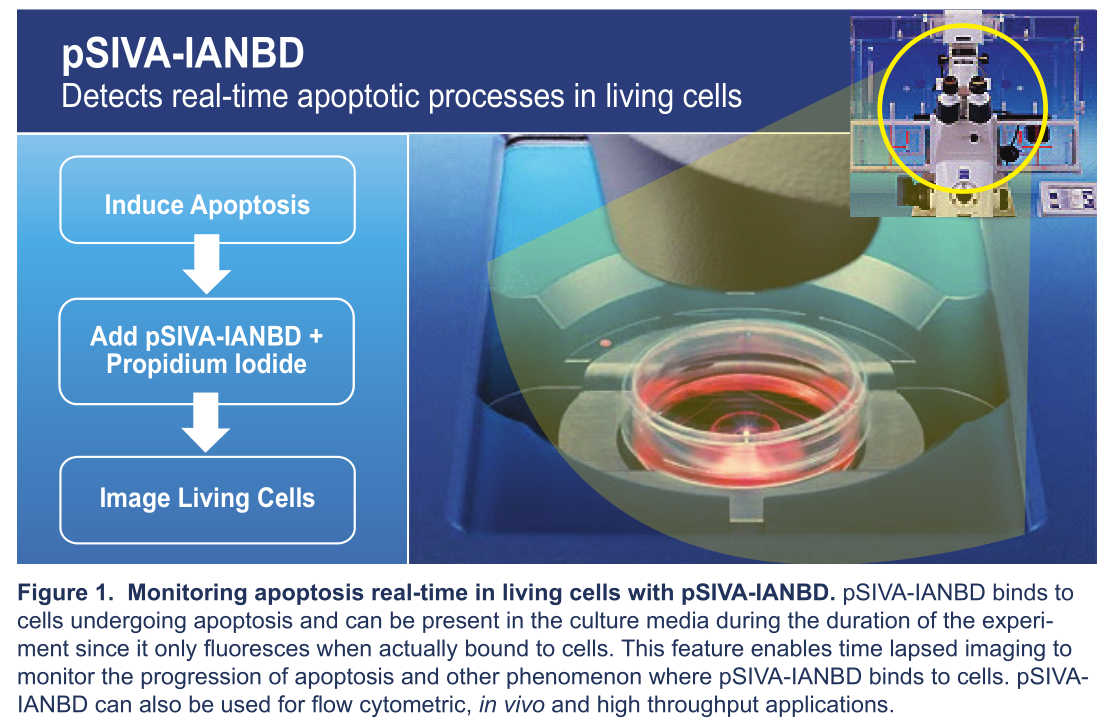
pSIVA(Polarity Sensitive Indicator of Viability & Apoptosis)凋亡檢測技術是Novus 2010年推出的一項新型技術,并于2013年發布**。該技術使用的pSIVA是一種Ca2+依賴性蛋白,與磷酯酰絲氨酸(PS)有非常高的結合能力,其與IANBD極性染料偶聯后形成pSIVA-IANBD復合物,這種復合物只有結合PS才會產生熒光。如下圖所示:

而傳統的Annexin V檢測技術,無論是否與PS結合,熒光始終存在,無法區分PS的瞬時與不可逆外翻,因此,相比較Annexin V,pSIVA能夠做到讓細胞凋亡檢測更精準。

?
?
|
Novus pSIVA試劑盒讓凋亡檢測更快更精準更直觀! 作者的喜愛與熱捧,該試劑盒操作相當簡單,只需將 pSIVA-IANBD或pSIVA-IANBD+PI直接加入細胞或組織, 孵育后便可檢測,無需洗滌! |
 |
?
在流式應用方面,Novus pSIVA凋亡試劑盒與Annexin V相比有一樣優異的表現:

在熒光觀察方面,Novus pSIVA凋亡試劑盒比Annexin V表現更出色!

Novus pSIVA凋亡檢測試劑盒產品列表
| 廠商 | 目錄號 | 產品名稱 | 規格 | 目錄價 | 文獻 |
| Novus | H6-NBP2-29611-25Tests | Polarity Sensitive Indicator of Viability Flow Assay Kit |
25Tests | 1850 | 13 |
| Novus | H6-NBP2-29611-100Tests | Polarity Sensitive Indicator of Viability Flow Assay Kit |
100Tests | 3040 | 13 |
| Novus | H6-NBP2-29382-1Sample SizeKit |
Polarity Sensitive Indicator of Viability Apoptosis Microscopy Kit |
1 Sample Size Kit | 1770 | 22 |
| Novus | H6-NBP2-29382 | Polarity Sensitive Indicator of Viability Apoptosis Microscopy Kit |
1 Kit | 3040 | 22 |
Novus pSIVA凋亡檢測試劑盒部分引用文獻列表:
[1]Neuronal deletion of caspase 8 protects against brain injury in mouse models of controlled cortical impact and kainic acid-induced excitotox- icity. Krajewska M, Z You, J Rong, C Kress, X Huang, J Yang, T Kyoda, R Leyva, S Banares, Y Hu, C-H Sze, MJ Whalen, L Salemena, R Hakem, BP Head, JC Reed, S Krajewski. PLOS ONE 6(9): e24341. doi:10.1371/journal.pone.0024341 (2011). IF (mouse primary neuron cultures): Figs 2B, 3.Cells were labeled with pSIVA-IANBD for 15 min, fixed and stained with a MAP2 antibody and DAPI. Cells double-stained with both pSIVA-IANBD and MAP2 (neuronal marker) were identified as degenerating neurons.
[2] Hostile takeover by Plasmodium: reorganization of parasite and host cell membranes during liver stage egress. Graewe S, KE Rankin, C Lehmann, C Deschermeier, L Hecht, U Froehlke, RR Stanway, V Heussler. PLOS ONE 7(9): e1002224 doi:10.1371/journal.ppat.1002224 (2011). IF (HepG2 cells infected with P. berghei parasites), Fig 1
[3] Mitochondrial NDUFS3 regulates the ROS-mediated onset of metabolic switch in transformed cells. Suhane S, H Kanzaki, V Arumugaswami, R Murali, VK Ramanujan. Biology Open doi: 10.1242/bio.20133244 (2013).pSIVA-IANBD Flow Kit: Flow (Cell Surface): Fig 1 (HEK293 cells). pSI- VA-IANBD was used to determine the basal level of apoptosis in HEK cells.
[4] Monitoring apoptosis and neuronal degeneration by real-time detection of phosphatidylserine externalization using a polarity-sensitive indica- tor of viability and apoptosis. Kim YE, J Chen, R Langen, JR Chan. Nature Protocols 5:1396-1405 (2010b). Time lapse microscopy of neurons innormal survival conditions and after NGF deprivation (Fig 2).
[5] A compact B model of huntingtin toxicity. Zhang CQ, Yeh T-l, A Leyva,LG Frank, J Miller, YE Kim, R Langen, S Finkbeiner, ML Amzel, CA Ross, MA Poirier. JBC 286:8188-8196 (2011). A pSIVA-IANBD based cell suspen- sion toxicity assay was used to determine cell viability in mouse Neu- ro2A (neuroblastoma) overexpressing huntingtin proteins (Fig 4).32 NBP2-29611 & NBP2-29382 pSIVA TM Kit pSIVA TM Kit Manual
[6] Diurnal, localized exposure of phosphatidylserine by rod outer seg-ment tips in wild-type but not Itgb5-/- or Mfge8-/- mouse retina. RuggieroL, MP Connor, J Chen, R Langen, SC Finnemann. PNAS 109:8145-4148(2012). Live tissue imaging (mouse retina): Figs 4, 5. S4. pSIVA-IANBDwas added to dissected live mouse retina and shown to label the tips of photoreceptor outer segments (POS).The results suggested that phosphatidylserine (PS) exposure is specif-ic to the POS surface. Furthermore, enhanced PS exposure preceded rod shedding and phagocytosis, suggesting that surface PS exposure promotes these processes.
[7]Characterization of cell cycle modifications induced by electric pulses in human corneal endothelium. Thi MH, Z He, N Campolmi, S Piselli, P Gain, M Peoc’H, JM Dumolllard, S Acquart, O Garraud, G Thuret. Acta Op- thalmologica 90:s249 (2012). Live tissue imaging (human cornea organ cultures): pSIVA-IANBD was used to assess cell death following gene electrotransfer into corneal endothelial cells.
[8.]Cell death goes LIVE: Technological advances in real-time tracking of cell death. Skommer J, Z Darzynkiewicz, D Wlodkowic. Cell Cycle 9:2330- 2341 (2010). Live cell imaging (etoposide treated cell lines & NGF-de-prived primary neuronal cultures): Discussion about tools for tracking cell death real-time.
[9]Annexin B12 Cys101,Cys260-N,N’-dimethyl-N-(iodoacetyl)-N’-(7-nitro-benz-2-oxa-1,3-diazol-4-yl)ethylenediamine. Shan L In: Molecular Imaging and Contrast Agent Database (MICAD) [Internet]. Bethesda (MD): National Center for Biotechnology Information (US); 2004-2013. http://www.ncbi.nlm.nih.gov/books/NBK45028/ (2010) Live cell imaging: The story of pSIVA as a phosphatidylserine-targeted, real-time, apoptosis-detecting probe.
[10] The change of cellular membranes on apoptosis: Fluorescence detection. Demchenko AP. Exp Oncol 34:263-268 (2012). Live imaging: Discussion about pSIVA as an important advancement in annexin based methodology.
[11]Real-time flow cytometry for the kinetic analysis of oncosis. Warnes G,S Martins. Cytometry Part A 79A:181-191 (2011). Live imaging: Discussion about pSIVA as an assay for real-time detection of apoptosis.
[12] Engineering a polarity-sensitive biosensor for time-lapse imaging of apoptotic processess and degeneration. Kim YE, J Chen, JR Chan, R Langen. Nature Methods 7:67-73 (2010a). Real-time live-cell imagingand time-lapse microscopy of apoptosis: Fig 2 (COS-7 cells), Fig 3 (rat33 NBP2-29611 & NBP2-29382 pSIVA TM KitpSIVA TM Kit Manual neuronal degeneration), Fig 4 (rat axonal degeneration), Fig 5 (rescue of rat neuronal degeneration as visualized by pSIVA.
[13]Monitoring apoptosis and neuronal degeneraton by real-time detection of posphatidylserine externalization using a polarity-sensitive indicator of viability and apoptosis. Kim YE, J Chen, R Langen, JR Chan. Nature Protocols 5:1396-1405 (2010b). Time lapse microscopy of neurons innormal survival conditions and after NGF deprivation (Fig 2).
[14] A compact beta model of huntingtin toxicity. Zhang CQ, Yeh T-I, A Leyva,LG Frank, J Miller, YE Kim, R Langen, S Finkbeiner, ML Amzel, CA Ross, MA Poirier. JBC 286:8188-8196 (2011). A pSIVA-IANBD based cell suspen- sion toxicity assay was used to determine cell viability in a mouse Neuro2A neuroblastoma cell line overexpressing huntingtin proteins (Fig 4).
[15]Hostile takeover by Plasmodium: reorganization of parasite and host cell membranes during liver stage egress. Graewe S, KE Rankin, CLehmann, C Deschermeier, L Hecht, U Froehike, RR Stanway, V Heussler. PLOS ONE 7(9): e1002224 doi:10.1371/journal.ppat.1002224 (2011). IF (HepG2 cells infected with P. berghei parasites), Fig 1.
[16] Novel applications of pSIVA, a new polarity sensitive phosphatidylser-ine (PS) apoptosis/cell death probe. Stein LS, PD Quintel, S Krajewski,JS Rosenberg, SK Singh. Cancer Res 72:237 (2012). Flow (Cell Surface):Jurkat (Figs 1-4). IF: Mouse primary neuronal cultures (Figs 5-6).
[17] Diurnal photoreceptor outer segment shedding: contributions of the RPE. Finnemann SC, L Ruggiero, MP Connor. Invest Opthalmol Vis Sci 53:4327 (2012). Live Imaging: mouse retina. pSIVA was used quantify the surface exposure of phosphotidylserine on the distal rod photore- ceptor outer segments.
[18] Flavopiridol synergizes with sorafenib to induce cytotoxicity and potentiate antitumorigenic activity in EGFR/HER-2 and mutant RAS/RAF breast cancer model systems. Nagaria TS, JL Williams, C Leduc, JA Squire, PA Greer, W Sangrar. Neoplasia 15:939-951 (2013). Flow cytometry (Cell surface): MDA-MB-231 (Fig 4A) and MDA-MB-468 (Fig 4B) adenocarcinoma cells.